Asian Spine J Search

CLOSE


Asian Spine J > Volume 17(6); 2023 > Article
Li, Li, Luk, Zhang, Sun, and Wang: Relationship between Fusion Mass Shift and Postoperative Distal Adding-on in Lenke 1 Adolescent Idiopathic Scoliosis after Selective Thoracic Fusion

Abstract

Study Design

This is a retrospective cohort study.

Purpose

This study aims to investigate the risk factors for postoperative distal adding-on in Lenke 1 adolescent idiopathic scoliosis (AIS) and validate the relationship between fusion mass shift (FMS) and postoperative distal adding-on.

Overview of Literature

Postoperative distal curve adding-on is one of the complications in AIS. FMS has been proposed to prevent postoperative distal adding-on, which requires further validation from different institutions.

Methods

This study included 60 patients with Lenke 1 AIS who underwent selective thoracic fusion surgery. Coronal spinal alignment parameters were analyzed preoperatively, postoperatively, and at the final follow-up. The postoperative FMS was divided into two groups: the balanced group (FMS ≤20 mm) and the unbalanced group (FMS >20 mm). An independent t-test was used to compare quantitative data between groups, and a chi-square test was used for qualitative data. Furthermore, binary logistic regression and receiver operating characteristics curve analyses were used to identify the risk factors for postoperative distal adding-on in AIS.

Results

At 2-year follow-up, the unbalanced group was more likely to have adding-on (17 of 24 patients) than the balanced group (six of 36 patients; p<0.001). Twenty-three patients with distal adding-on had significantly greater preoperative and postoperative lower instrumented vertebrae (LIV) rotation, FMS, and FMS angle (FMSA) than those without postoperative distal adding-on. Binary logistic regression analysis selected three independent risk factors for adding-on incidence after surgery: FMS (odds ratio [OR], 1.115; 95% confidence interval [CI], 1.049–1.185; p<0.001), FMSA (OR, 1.590; 95% CI, 1.225–2.064; p<0.001), and postoperative LIV rotation (OR, 6.581; 95% CI, 2.280–19.000; p<0.001).

Conclusions

Achieving a balanced fusion mass intraoperatively is important to avoid postoperative distal adding-on, with FMS of <20 mm and FMS angle of <4.5°. Furthermore, correcting LIV rotation helps to decrease the incidence of postoperative distal adding-on.

Introduction

The etiology of adolescent idiopathic scoliosis (AIS), which affects 1%–3% of adolescents, remains unknown [1]. The incidence of Lenke type 1 scoliosis accounts for 51% of AIS [2]. Selected thoracic fusions (STFs) are performed on these patients to achieve coronal and sagittal balance and to preserve more lumbar motion segments. Postoperative distal adding-on is one of the complications after STF in Lenke 1 AIS.
Postoperative distal adding-on is defined as a progressive increase in the number of vertebrae included distally in the primary curvature, combined with a deviation of <5 mm from the center sacral vertical line (CSVL) to the lower instrumented vertebrae (LIV) or angulation of the first disk caudal to the LIV of more than 5° [3]. The occurrence of postoperative distal adding-on is closely related to decreased postoperative satisfaction, back pain, increased cost, and reoperation [4,5].
In 2017, the concept of fusion mass shift (FMS) was proposed, with an intraoperative aim of balanced fusion mass with FMS of <20 mm to avoid postoperative distal adding-on [3]. Therefore, we retrospectively reviewed the Lenke 1 AIS cases from the last decade to validate the influence of FMS on the occurrence of postoperative distal adding-on and to investigate the risk factors for postoperative distal adding-on in Lenke 1 AIS.

Materials and Methods

This is a retrospective study of patients from a single institution. Permission to conduct this retrospective study was obtained from the ethics committee of Shandong Provincial Hospital Affiliated to Shandong First Medical University (IRB approval no., NSFC-2020-528), and written informed consent was obtained from all subjects who participated in this study.
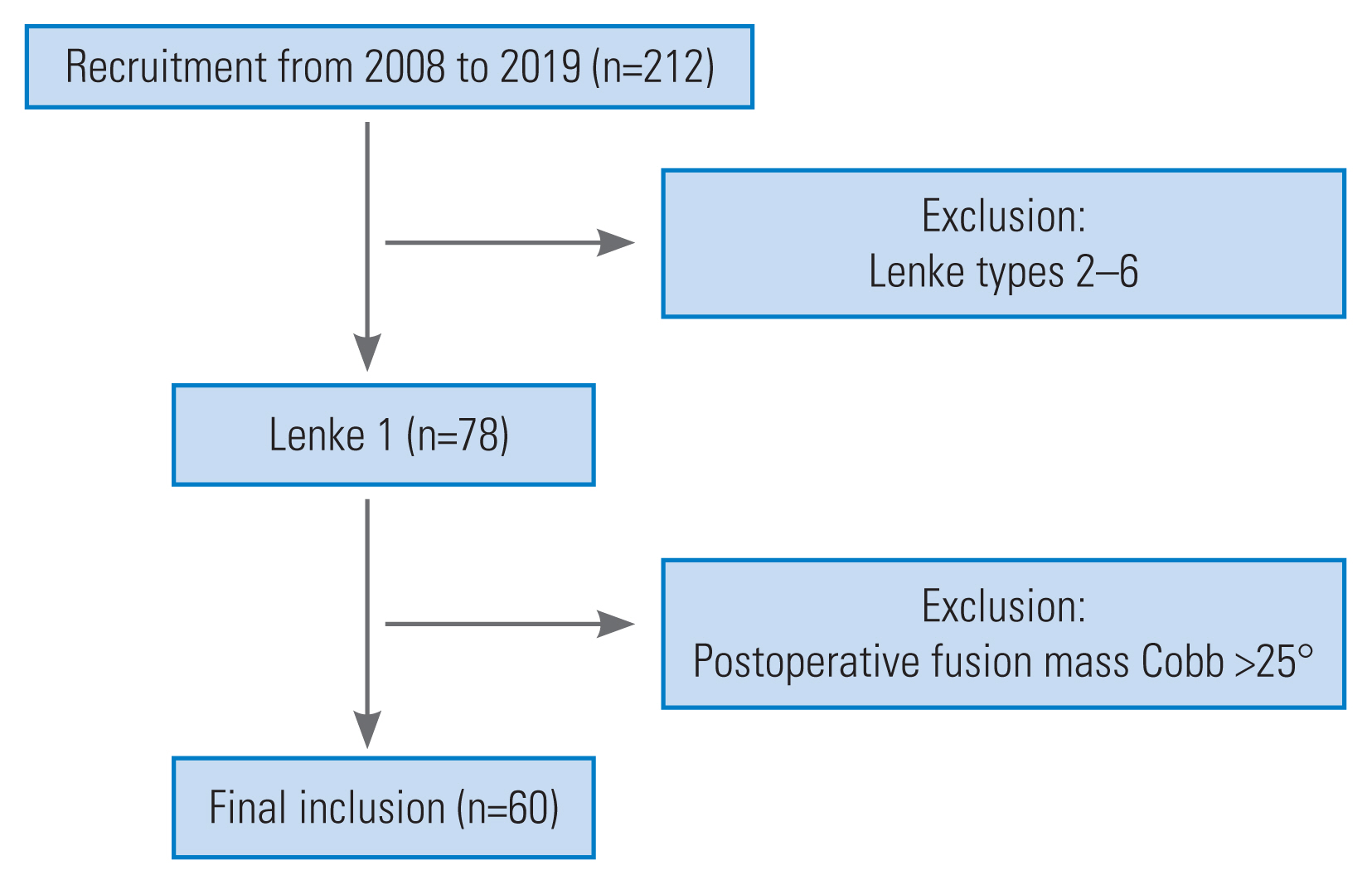
The study included 78 patients with Lenke 1 AIS who underwent selective thoracic fusion surgery between 2008 and 2019. Of them, 60 patients were included in the study, and 18 were excluded (Fig. 1). They all underwent surgical treatment by the same surgeon in a university-affiliated hospital. The fusion levels for selective thoracic fusion in Lenke 1 AIS were assessed using the fulcrum bending radiograph, a method described by Luk et al. [6].
The inclusion criteria of this study are as follows: (1) patients aged between 10 and 18 years, (2) patients with AIS who have main thoracic (MT) scoliosis, (3) the surgical procedure is selective thoracic fusion, (4) fixation with pedicle screw system, and (5) patients with a minimum of 2 years of follow-up.
The exclusion criteria of this study are as follows: (1) patients with other types of AIS, (2) patients with other types of scoliosis, (3) patients with a prior history of spinal surgery, (4) patients with a history of spinal infection, and (5) patients with postoperative fusion mass Cobb (FMC) angle of more than 25°.
Coronal spinal alignment parameters on standing anteroposterior (AP) plain radiographs were analyzed preoperatively, postoperatively, and at the final follow-up according to the established positioning protocol. The coronal spinal alignment parameters included the proximal thoracic (PT) curve, MT curve, thoracolumbar/lumbar (TL/L) curve, clavicle angle (CA), thoracic trunk shift (TTS), T1 tilt, LIV rotation, FMC angle (Cobb angle between the superior endplate of the upper instrumented vertebrae and the inferior endplate of LIV), FMS (distance from the center of the superior endplate of upper instrumented vertebrae to a perpendicular line of the inferior endplate of LIV erected from the center of the LIV), FMSA (angle between a line from the center of the superior endplate of upper instrumented vertebrae to the center of the inferior endplate of LIV and a perpendicular line of the inferior endplate of LIV) (Fig. 2). FMC, FMS, and FMSA were measured on the postoperative films. In CA and T1 tilt, the left higher was positive, and the right higher was negative. Furthermore, age, sex, triradiated cartilage, and Risser sign were recorded before surgery. The Scoliosis Research Society 22-item patient questionnaire (SRS-22) was used to evaluate clinical improvement preoperatively, postoperatively, and at the final follow-up [7].
The distal adding-on phenomenon was assessed by comparing postoperative standing AP radiographs with final follow-up standing AP radiographs. The postoperative FMS was divided into two groups: the balanced group (FMS ≤20 mm) and the unbalanced group (FMS >20 mm).
Statistical analysis was analyzed using the IBM SPSS ver. 27.0 software (IBM Corp., Armonk, NY, USA). An independent t-test was used to compare quantitative data between groups, and a chi-square test was used for qualitative data. In addition, binary logistic regression and receiver operating characteristics (ROC) curve analyses were used to identify the risk factors for postoperative distal adding-on in Lenke 1 AIS. The distribution of parameters was presented as mean and standard deviation. The significance level was set to p<0.05.

Results

This study included 60 patients (13 males and 47 females). The mean age for surgery was 13.6 years old, with a 2-year follow-up. Twenty-three patients developed complications of distal adding-on at 2-year follow-up. Significant differences in preoperative parameters were identified between no adding-on and adding-on patients in trunk shift (13.84±14.71 mm versus 20.51±10.86 mm) and LIV rotation (0.57±0.69 grade versus 1.09±0.79 grade). In addition, significant differences in parameters were observed postoperatively between no adding-on and adding-on groups in the TL/L curve (9.97°±9.75° versus 14.9°±8.25°), LIV rotation (0.35±0.59 grade versus 1.13±0.81 grade), FMS (12.13±9.27 mm versus 27.15±14.65 mm), and FMSA (2.73°±2.1° versus 6.38°±3.48°). At the final follow-up, there were significant differences in parameters between no adding-on and adding-on groups in the TL/L curve (10.63°±8.52° versus 15.75°±10.49°) and LIV rotation (0.35±0.59 versus 1.09±0.79) (Table 1).
Age, gender, Risser sign, and preoperative and postoperative coronal spinal alignment parameters did not differ between balanced and unbalanced groups. On the other hand, patients in the unbalanced group had significantly lower mental health scores than those in the balanced group (Tables 2, 3). In addition, the TL/L curve showed significant differences between balanced and unbalanced groups at 2-year follow-up (10±8 versus 16±10), which was correlated with the incidence rate of the distal adding-on phenomenon.
Significant differences in FMC (11°±6° versus 19°±9°), FMS (11±6 mm versus 32±9 mm), and FMSA (2°±2° versus 7°±3°) were observed between balanced and unbalanced groups. Even though there were no significant differences in preoperative and postoperative LIV rotation between groups, postoperative LIV rotation was higher in the unbalanced group than preoperative LIV rotation (1.0±0.9 grade versus 0.7±0.9 grade). In addition, the unbalanced group was more likely to develop distal adding-on at 2-year follow-up (17 of 24 patients) than the balanced group (six of 36 patients) (Table 2).
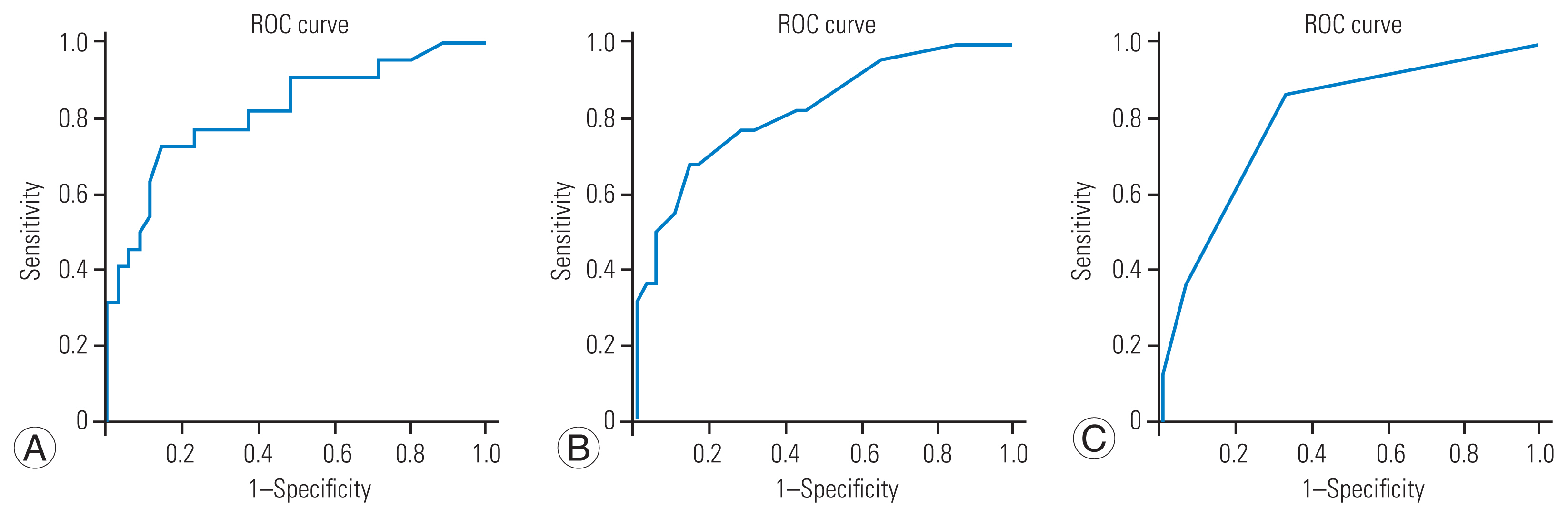
Binary logistic regression analysis revealed three independent risk factors for distal adding-on incidence after surgery: FMS (odds ratio [OR], 1.115; 95% confidence interval [CI], 1.049–1.185; p<0.001), FMSA (OR, 1.590; 95% CI, 1.225–2.064; p<0.001), and postoperative LIV rotation (OR, 6.581; 95% CI, 2.280–19.000; p<0.001) (Table 4). ROC curve analysis confirmed the following risk factors: area under the ROC curve (AUC) of FMS was 0.831 (p<0.001) with an optimal cutoff value of 20°, AUC of FMSA was 0.856 (p<0.001) with an optimal cutoff value at 4.5°, and AUC of postoperative LIV rotation was 0.812 (p=0.031) with an optimal cutoff value of 1 (Fig. 3).

Discussion

Postoperative distal curve adding-on is a complication of AIS undergoing selective thoracic fusion surgery. In our study, the incidence of postoperative distal adding-on was 38.3%, similar to the previous study (21%–51%) [3,8]. Previous studies reported that a low Risser grade predicted a high growth potential and was more likely to develop distal adding-on after selected thoracic fusion surgery [9,10]. The selection of LIV was another important factor influencing the incidence of postoperative distal adding-on [8,11]. According to He et al. [12], preoperative rotation of LIV was also an independent predictor of postoperative distal adding-on in Lenke 1A or 2A AIS patients.
In 2017, the concept of balanced fusion mass was proposed [3], which helped to reduce postoperative distal adding-on. In the present study, a distal adding-on phenomenon was observed in 16.7% (six of 36 patients) of the balanced group (FMS ≤20 mm) and 70.8% (17 of 24 patients) of the unbalanced group (FMS >20 mm). This was consistent with previous results, which showed that 12.2% (five of 41 patients) were in the balanced group, and 54.5% (six of 11 patients) were in the unbalanced group [3]. Therefore, the aim of corrective surgery should be to achieve a balanced fusion mass. This should be done intraoperatively to reduce the risk of postoperative distal adding-on. FMS, FMSA, and rotation of LIV are parameters that determine whether a balanced fusion mass is achieved.
Aside from FMS, FMS angle was found to be an independent risk factor for postoperative distal adding-on. FMS could help us preoperatively determine fusion levels through fulcrum bending radiographs [6]. We can also use FMS and FMSA during the surgery to assess whether the balanced fusion mass is achieved. However, intraoperative fluoroscopy was typically performed using a C-arm X-ray machine, and measuring the FMS without scale was challenging. FMSA, which is highly correlated with FMS, is easier to be measured in fluorography without scale. Furthermore, intraoperative FMS is not weight-bearing, and FMSA may be more sensitive than FMS in short fusion in Lenke 1 AIS patients undergoing selected thoracic fusion surgery. In our study, avoiding a residual FMSA of more than 4.5° could help reduce the incidence of the postoperative distal adding-on phenomenon.
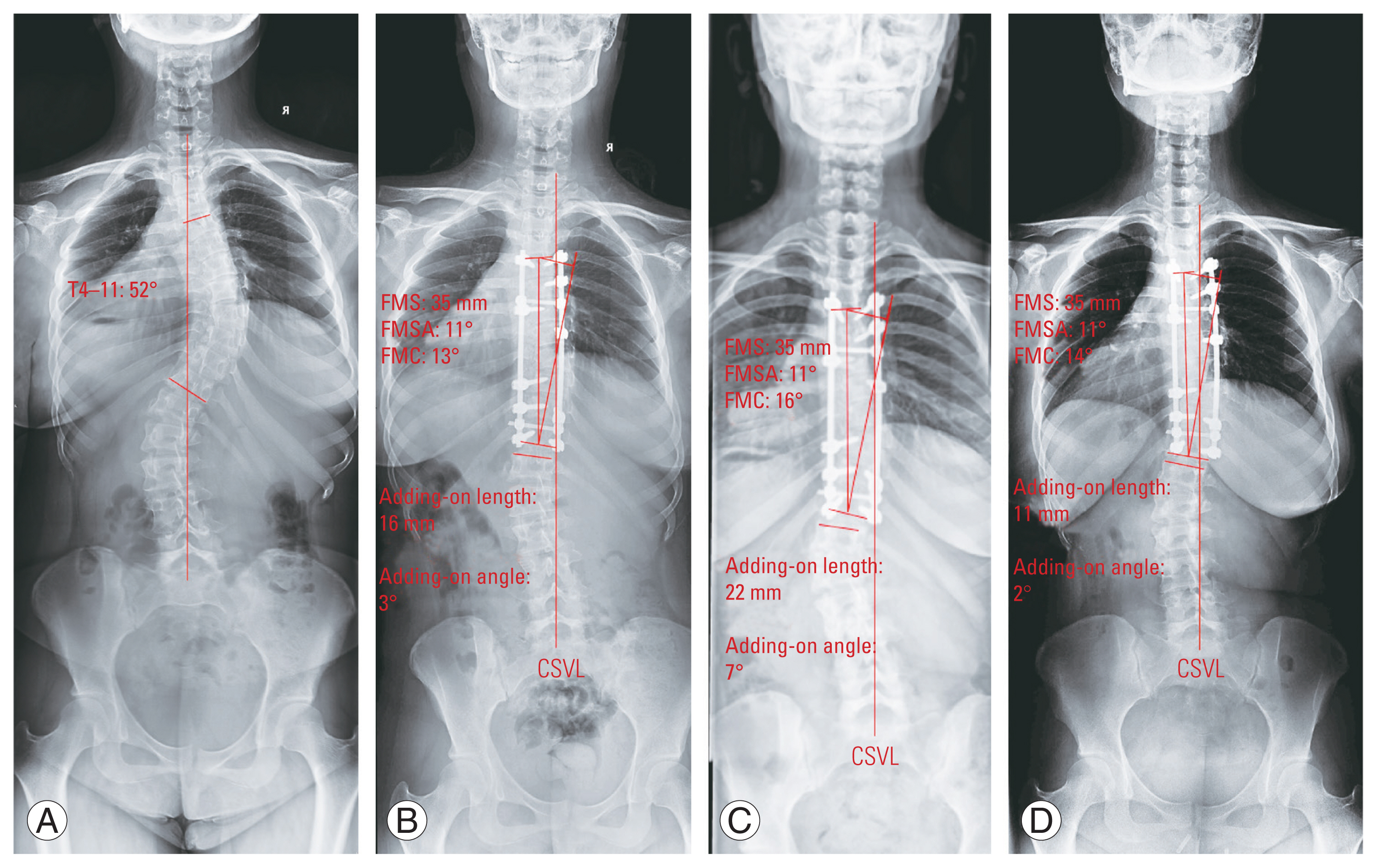
FMS occurred before the postoperative distal adding-on phenomenon. It occurred during the operation at the completion of the fixation and remained unchanged after the operation. However, the postoperative distal adding-on phenomenon gradually developed at serial postoperative follow-up X-rays (Fig. 4). In our study, an FMS of >20 mm was one of the causes of the postoperative distal adding-on phenomenon.
FMS can be assessed intraoperatively using a long cross-metal bar perpendicular to the lower endplate of LIV. It facilitates the assessment of the fusion mass. However, because this assessment is performed in a nonweight-bearing position, the unfused segments can further compensate postoperatively when the patient assumes an erect posture. Therefore, a postoperative adding-on phenomenon will occur in the presence of a large residual FMS or FMSA.
The postoperative LIV rotation was also an important risk factor contributing to postoperative distal adding-on in Lenke 1 AIS patients. The residual rotation could cause a distal adjacent segment to be offset. The concept of neutral vertebrae was proposed by Suk et al. [13], who suggested that it should be fused to neutral vertebrae when neutral vertebrae were two vertebrae caudal to lower end vertebrae. The selection of LIV should take into account the vertebrae rotation. Correction of LIV rotation may be beneficial in reducing distal adding-on.
A previous study reported that postoperative shoulder imbalance had significantly associated with distal adding-on in Lenke 2 AIS patients [14]. However, in our study, postoperative shoulder imbalance was not an independent risk factor for distal adding-on in Lenke 1 AIS. The PT curve was the main compensatory to balance the postoperative shoulder. Although postoperative CA was not significantly associated with distal adding-on in the present study, it may compensate for postoperative shoulder imbalance [15].
In our study, no patients with postoperative distal adding-on had a second surgery. In most cases, the truncal shift with distal adding-on phenomenon could be well controlled after strengthening the back muscles, as shown in Fig. 4. She was a 14-year-old female with a 52° MT preoperatively. After surgery, the MT was well corrected but with a large FMS and FMSA. The distal adding-on phenomenon occurred at the 6-month follow-up, with a deviation of 6 mm from the CSVL to the LIV. After strengthening the lumbar and back muscles, the adding-on phenomenon is controlled and does not require revision surgery at a 4-year follow-up.
This study has some limitations. First, the sample size is small due to the single-institution research design. A larger sample of multicenter studies is being conducted. Second, only Lenke 1 AIS was included in the study. Third, this study did not analyze whether sagittal lumbar alignment parameters influence the adding-on.

Conclusions

Distal adding-on phenomenon occurred in 38.3% of patients. Therefore, achieving a balanced fusion mass intraoperatively was important to avoid postoperative distal adding-on, with FMS of <20 mm and FMS angle of <4.5°. Furthermore, correcting LIV rotation helped decrease the incidence of postoperative distal adding-on.

Notes

Conflict of interest

No potential conflict of interest relevant to this article was reported.

Funding

This article receives funding from Natural Science Foundation of Shandong Province (award number: ZR2020QH264) and Clinical Medical Science and Technology Innovation Plan of Jinan (award number: 202019202).

Author Contributions

Jianlong Li and Jianmin Sun did the data collection and analysis. Yang Li and Chenggui Zhang finished the measurement. Keith D. K. Luk did the critical revision. Guodong Wang designed the study and wrote the article.

Fig. 1
A flowchart shows the recruitment process of patients in this study.
asj-2022-0466f1.jpg
Fig. 2
The graph shows the measurement guide of coronal spinal alignment parameters. (A) Clavicle angle (CA) represents the angle between the line connecting the highest points of the clavicles and the horizontal plane, and T1 tilt represents the angle between the upper endplate of T1 vertebra with the horizontal plane. (B) Thoracic trunk shift (TTS) represents the distance between center sacral vertical line (CSVL) and the midpoint of the two widest points of the rib cage along a horizontal parallel line. (C) Fusion mass shift (FMS) and fusion mass shift angle (FMSA) is measured in fluorography intraoperatively.
asj-2022-0466f2.jpg
Fig. 3
The graph shows the result of receiver operating characteristics (ROC) curve analysis for risk factors of distal adding-on: fusion mass shift (A), fusion mass shift angle (B), and postoperative lower instrumented vertebrae rotation (C).
asj-2022-0466f3.jpg
Fig. 4
(A) The graph shows a 14-year-old girl with a 52° main thoracic preoperatively. (B) She underwent a selective thoracic fusion with fusion mass Cobb (FMC) of 13°, but has fusion mass shift (FMS) of 35 mm and fusion mass shift angle (FMSA) of 11°. (C) The distal adding-on phenomenon was occurred at 6-month follow-up, which with an increase of 6 mm deviating from the center sacral vertical line (CSVL) to lower instrumented vertebrae (LIV) and an increase of 4° in angulation of the first disc caudal to the LIV. (D) At 4-year follow-up, the adding-on phenomenon improved better than 6-month follow-up.
asj-2022-0466f4.jpg
Table 1
Comparison of coronal spinal alignment parameters between non-PJK and PJK group
Variable No adding-on group (n=37) Adding-on group (n=23) p-value
PT (°)
 Preoperative 29.97±9.25 31.43±12.07 0.6
 Postoperative 20.06±9.1 17.98±10.89 0.43
 Final follow-up 18.37±9.08 16.16±9.09 0.37
MT (°)
 Preoperative 56.06±14.3 58.4±12.06 0.51
 Postoperative 20.32±14.68 18.32±9.29 0.52
 Final follow-up 10.63±8.52 18.84±10.04 0.71
TL/L (°)
 Preoperative 33.5±10.23 35.88±9.42 0.37
 Postoperative 9.97±9.75 14.9±8.25 0.02*
 Final follow-up 10.63±8.52 15.75±10.49 0.04*
Clavicle angle (°)
 Preoperative −2.16±2.5 −1.26±2.6 0.42
 Postoperative 1.42±2.15 2.35±3.34 0.25
 Final follow-up 1.31±2.15 1.14±1.9 0.75
Trunk shift (mm)
 Preoperative 13.84±14.71 20.51±10.86 0.05*
 Postoperative −6.16±9.97 −6±10.42 0.95
 Final follow-up −4.24±7.95 −4.75±11.79 0.85
T1 tilt (°)
 Preoperative 0.01±5.59 0.7±4.38 0.62
 Postoperative 4.28±4.18 4.11±5.21 0.89
 Final follow-up 3.73±4.44 5.05±5.43 0.36
LIV rotation
 Preoperative 0.57±0.69 1.09±0.79 0.01*
 Postoperative 0.35±0.59 1.13±0.81 <0.01*
 Final follow-up 0.35±0.59 1.09±0.79 <0.01*
Postoperative FMC (°) 12.36±8.38 16.7±8.38 0.06
Postoperative FMS (mm) 12.13±9.27 27.15±14.65 <0.01*
Postoperative FMSA (°) 2.73±2.1 6.38±3.48 <0.01*

Values are presented as mean±standard deviation.

PJK, proximal junctional kyphosis; PT, proximal thoracic curve; MT, major thoracic curve; TL/L, thoracolumbar/lumbar curve; LIV, lower instrumented vertebrae; FMC, fusion mass Cobb angle; FMS, fusion mass shift; FMSA, fusion mass shift angle.

* p<0.05 (statistically significant).

Table 2
Comparison of demographic and clinical information between the balanced and unbalanced group
Variable Balanced group Unbalanced group p-value
Gender 0.536
 Male 7 6
 Female 29 18
Age (yr) 13.4±1.5 13.8±1.7 0.423
Risser sign 0.213
 0 6 2
 1 5 3
 2 1 4
 3 4 4
 4 20 9
 5 0 2
Preoperative SRS-22 outcome
 Function 4.461±0.278 4.575±0.259 0.111
 Pain 4.711±0.175 4.700±0.213 0.833
 Self-image 3.594±0.343 3.642±0.306 0.580
 Mental health 4.217±0.353 3.908±0.425 0.005*
 Satisfaction 4.583±0.604 4.667±0.637 0.615
 Total 4.639±0.683 4.625±0.576 0.933
Final follow-up SRS-22 outcome
 Function 4.461±0.278 4.575±0.2593 0.116
 Pain 4.711±0.175 4.700±0.213 0.826
 Self-image 3.594±0.343 3.642±0.306 0.588
 Mental health 4.217±0.353 3.908±0.425 0.003*
Satisfaction 4.583±0.604 4.667±0.637 0.610
 Total 4.639±0.683 4.625±0.576 0.935
FMC 11±6 19±9 <0.001*
FMS 11±6 32±9 <0.001*
FMSA 2±2 7±3 <0.001*
UIV location
 T3 or 4 21 16
 T5 or 6 14 6 0.149
LIV location
 T11 or 12 10 4
 L1 or 2 or 3 25 18 0.285
Distal adding on
 Absence 30 7
 Presence 6 17 <0.001*

Values are presented as number or mean±standard deviation.

SRS-22, Scoliosis Research Society 22-item patient questionnaire; FMC, fusion mass Cobb; FMS, fusion mass shift; FMSA, fusion mass shift angle; UIV, upper instrumented vertebrae; LIV, lower instrumented vertebrae.

* p<0.05 (statistically significant).

Table 3
Comparison of radiographic parameter between the balanced and unbalanced groups
Variable Balanced group Unbalanced group p-value
Preoperative parameters
 PT (°) 30±11 33±11 0.316
 MT (°) 54±14 61±12 0.066
 TL/L (°) 33±10 39±9 0.262
 CA (°) −2±2 −2±2 0.735
 TTS (mm) 19±13 14±14 0.121
 T1 tilt (°) 0±6 0±5 0.802
 LIV rotation 0.9±0.7 0.7±0.9 0.420
Postoperative parameters
 PT (°) 18±9 22±10 0.115
 MT (°) 18±15 22±9 0.208
 TL/L (°) 15±9 10±7 0.169
 CA (°) 1±2 2±3 0.739
 TTS (mm) −6±11 −6±9 0.885
 T1 tilt (°) 5±6 4±4 0.631
 LIV rotation 0.6±0.8 1.0±0.9 0.055
Final follow-up parameters
 PT (°) 16±9 20±9 0.390
 MT (°) 17±14 23±9 0.097
 TL/L (°) 10±8 16±10 0.009*
 CA (°) 1±2 1±2 0.970
 TTS (mm) −5±11 −4±8 0.535
 T1 tilt (°) 6±6 3±4 0.058
 LIV rotation 0.5±0.7 0.9±0.8 0.092

Values are presented as number or mean±standard deviation.

PT, proximal thoracic; MT, main thoracic; TL/L, thoracolumbar/lumbar; CA, clavicle angle; TTS, thoracic trunk shift; LIV, lower instrumented vertebrae.

* p<0.05 (statistically significant).

Table 4
Binary logistic regression analysis of risk factors for distal adding on after correction surgery of Lenke 1 adolescent idiopathic scoliosis
Variable OR (95% CI) p-value
FMS 1.115 (1.049–1.185) <0.001*
FMSA 1.590 (1.225–2.064) <0.001*
FMC 1.040 (0.973–1.111) 0.251
CA preop 1.024 (0.823–1.273) 0.834
CA postop 1.234 (0.963–1.582) 0.096
CA f/u 0.855 (0.641–1.140) 0.285
Shoulder imbalance 0.425 (0.111–1.632) 0.213
Age 0.897 (0.629–1.280) 0.550
Risser sign 1.063 (0.740–1.521) 0.654
UIV location 1.025 (0.662–1.585) 0.913
LIV location 0.700 (0.462–1.060) 0.092
LIV rotation preop 1.222 (0.626–2.384) 0.557
LIV rotation postop 6.581 (2.280–19.000) <0.001*
LIV rotation f/u 6.230 (2.251–17.245) <0.001*
MT correction rate 0.565 (0.009–36.596) 0.788

OR, odds ratio; CI, confidence interval; FMS, fusion mass shift; FMSA, fusion mass shift angle; FMC, fusion mass Cobb; CA, clavicle angle; preop, preoperative; postop, postoperative; f/u, follow-up; UIV, upper instrumented vertebrae; LIV, lower instrumented vertebrae; MT, main thoracic.

* p<0.05 (statistically significant).

References

1. Dunn J, Henrikson NB, Morrison CC, Blasi PR, Nguyen M, Lin JS. Screening for adolescent idiopathic scoliosis: evidence report and systematic review for the US Preventive Services Task Force. JAMA 2018;319:173–87.
crossref pmid
2. Lenke LG, Betz RR, Harms J, et al. Adolescent idiopathic scoliosis: a new classification to determine extent of spinal arthrodesis. J Bone Joint Surg Am 2001;83:1169–81.
pmid
3. Shigematsu H, Cheung JP, Bruzzone M, et al. Preventing Fusion mass shift avoids postoperative distal curve adding-on in adolescent idiopathic scoliosis. Clin Orthop Relat Res 2017;475:1448–60.
crossref pmid pmc pdf
4. Sponseller PD, Betz R, Newton PO, et al. Differences in curve behavior after fusion in adolescent idiopathic scoliosis patients with open triradiate cartilages. Spine (Phila Pa 1976) 2009;34:827–31.
crossref pmid
5. Behensky H, Cole AA, Freeman BJ, Grevitt MP, Mehdian HS, Webb JK. Fixed lumbar apical vertebral rotation predicts spinal decompensation in Lenke type 3C adolescent idiopathic scoliosis after selective posterior thoracic correction and fusion. Eur Spine J 2007;16:1570–8.
crossref pmid pmc pdf
6. Luk KD, Don AS, Chong CS, Wong YW, Cheung KM. Selection of fusion levels in adolescent idiopathic scoliosis using fulcrum bending prediction: a prospective study. Spine (Phila Pa 1976) 2008;33:2192–8.
pmid
7. Cheung KM, Senkoylu A, Alanay A, Genc Y, Lau S, Luk KD. Reliability and concurrent validity of the adapted Chinese version of Scoliosis Research Society-22 (SRS-22) questionnaire. Spine (Phila Pa 1976) 2007;32:1141–5.
crossref pmid
8. Wang Y, Hansen ES, Hoy K, Wu C, Bunger CE. Distal adding-on phenomenon in Lenke 1A scoliosis: risk factor identification and treatment strategy comparison. Spine (Phila Pa 1976) 2011;36:1113–22.
pmid
9. Sanders JO, Herring JA, Browne RH. Posterior arthrodesis and instrumentation in the immature (Risser-grade-0) spine in idiopathic scoliosis. J Bone Joint Surg Am 1995;77:39–45.
crossref pmid
10. Tan JH, Hey HW, Wong G, Wong HK, Lau LL. Lumbar adding-on of the thoracic spine after selective fusion in adolescent idiopathic scoliosis lenke types 1 and 2 patients: a critical appraisal. Spine (Phila Pa 1976) 2021;46:E167–73.
crossref pmid
11. Li Y, Bai H, Liu C, et al. Distal adding-on phenomenon in Lenke IA and Lenke IIA: risk analysis and selection of the lowest instrumented vertebra. World Neurosurg 2020;136:e171–80.
crossref pmid
12. He Z, Qin X, Yin R, et al. The rotation of preoperative-presumed lowest instrumented vertebra: is it a risk factor for distal adding-on in Lenke 1A/2A curve treated with selective thoracic fusion? Eur Spine J 2020;29:2054–63.
crossref pmid pdf
13. Suk SI, Lee SM, Chung ER, Kim JH, Kim SS. Selective thoracic fusion with segmental pedicle screw fixation in the treatment of thoracic idiopathic scoliosis: more than 5-year follow-up. Spine (Phila Pa 1976) 2005;30:1602–9.
pmid
14. Cao K, Watanabe K, Hosogane N, et al. Association of postoperative shoulder balance with adding-on in Lenke type II adolescent idiopathic scoliosis. Spine (Phila Pa 1976) 2014;39:E705–12.
crossref pmid
15. Matsumoto M, Watanabe K, Kawakami N, et al. Postoperative shoulder imbalance in Lenke type 1A adolescent idiopathic scoliosis and related factors. BMC Musculoskelet Disord 2014;15:366.
crossref pmid pmc pdf


ABOUT
ARTICLE CATEGORY

Browse all articles >

BROWSE ARTICLES
EDITORIAL POLICY
FOR CONTRIBUTORS
Editorial Office
Department of Orthopedic Surgery, Asan Medical Center, University of Ulsan College of Medicine
88, Olympic-ro 43-gil, Songpa-gu, Seoul 05505, Korea
Tel: +82-2-3010-3530    Fax: +82-2-3010-8555    E-mail: asianspinejournal@gmail.com                
Korean Society of Spine Surgery
27, Dongguk-ro, Ilsandong-gu, Goyang-si 10326, Korea
Tel: +82-31-966-3413    Fax: +82-2-831-3414    E-mail: office@spine.or.kr                

Copyright © 2024 by Korean Society of Spine Surgery.

Developed in M2PI

Close layer
prev next