Introduction
Intradural arachnoid cysts are uncommon and arise in thoracic, lumbar and cervical region in decreasing frequency. Arachnoid cyst are mostly located on the posterior aspect of the cord. This means that anteriorly located arachnoid cyst are exceptional, particularly those occurring in the cervical spinal region [1]. Regarding the rarity of this pathology in this certain location, it should be noted that eight out of nine cases of intradural arachnoid cyst reported by Kendall et al. [1] lied posteriorly in the thoracic region. Moreover, of 17 cases of intradural arachnoid cyst including eight cases of cervical region reported by Alvisi et al. [2], all were located posteriorly. A literature search by Kazan et al. [3] from 1974 to 1999 revealed only 10 cases of anterior cervical intradural arachnoid cyst including two cases of their own. However, apparently six cases escaped their attention [1,4]. In current article, besides very careful resurvey of the literature up to 1999, we updated the review yielding 24 cases reported in English literature [1,3-21]. Herein, two additional cases who have undergone surgery with good postoperative neural recovery are presented.
Case Report
1. Case 1
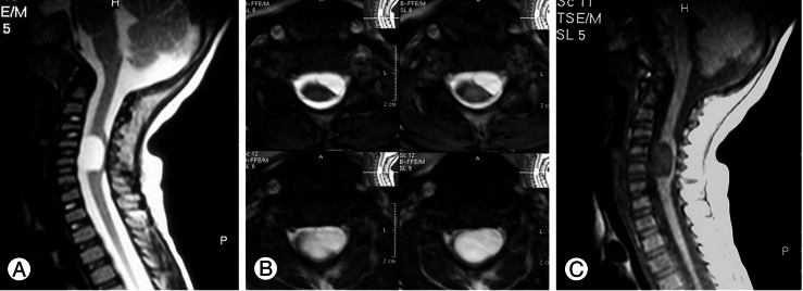
This 2-year-old girl was admitted to our hospital with a 2-week history of torticollis and weakness of the left upper extremity for a week. She had no history of trauma or infection of upper respiratory tract infection. Her neurological exam showed moderate paresis of the left upper extremity, all reflexes were hyperactive and plater reflex was extensor on the left side. Cervical spine X-Ray and open mouth view were regarded normal. Magnetic resonance imaging (MRI) revealed an intradural hypointense cystic mass anterior to the cord in T1 and hyperintense in T2 images resembling cerebrospinal fluid (CSF) intensity at C5-C6 level which was markedly compressing the cord (Fig. 1). A provisional diagnosis of intradural arachnoid cyst was made.
With the patient in prone position laminectomy of C5 and C6 was done. The dura was tense and on opening the cord was flattened and budged posteriorly into the dural incision. The dentate ligaments at C5-C6 level were cut on the left side revealing a translucent cystic collection anterior to the cord. The cyst was aspirated revealing CSF. Following aspiration, the cyst shrunk and the cord pulsation appeared. With gentle retraction of the cord, the cyst's capsule could be excised totally. Postoperatively, the child showed motor recovery and her neurological examination three months after surgery was normal.
2. Case 2
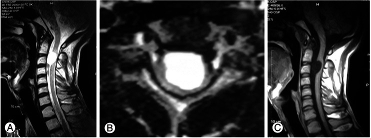
This 17-year-old male was referred with dull moderate neck pain followed with progressive weakness of the right upper extremity of one month duration followed by gait disturbances in recent days. On clinical survey, he was found to have quadriparesis most prominent in the right upper extremity. Cervical MRI disclosed a ventrally located intradural cystic lesion with apparent cord compression at C2-C3 level (Fig. 2).
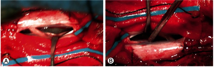
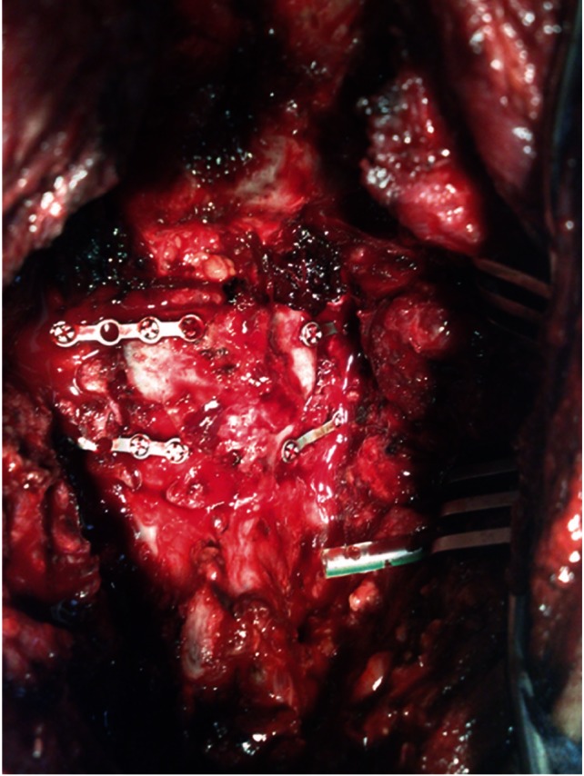
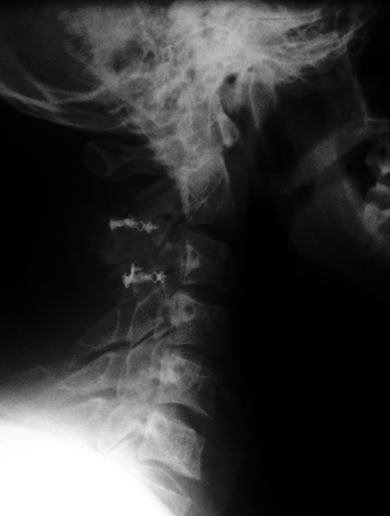
With the patient in prone position, a cervical midline incision was made at the appropriate levels. En-block laminotomy of C2 and C3, incising the corresponding laminas bilaterally with the aid of high speed drill was done. The dura seemed under considerable pressure. The techal sac was open in the midline. This revealed a splayed cord under considerable pressure from the anterior. After sectioning dentate ligament on the right side and mild retraction of the cord, a cystic mass with transparent wall became visible. The wall of the cyst was minimally incised disclosing clear content compatible with CSF. Subsequently, the cyst was widely fenestrated into the subarachnoid space with partial removal of the capsule (Fig. 3). The cord was decompressed and returned to its normal position with good pulsation. Later, after dural closure, two level laminoplasty with application of mini plates was done bilaterally (Figs. 4, 5). The patient made uneventful recovery and post operative cervical MRI showed resolution of the arachnoid cyst (Fig. 6). He returned to his previous activities within six months.
Discussion
Intradural arachnoid cysts are uncommon and most of them are located in the thoracic region followed with the lumbar and the cervical region [1,2]. Majority of these cysts lie posteriorly on the spinal cord and are rarely located anteriorly. Review of the literature revealed that anteriorly located intradural arachnoid cyst are very rare in the cervical region and we could find only 24 examples reported previously. With taking to account two current cases, the number of the cases published so far in the literature reach to 26 cases (Table 1).
Of the total these 26 reviewed cases, 15 occurred in males and the remaining 11 were female. Age ranged from 2 to 43 years with mean of 15/1 years. In 19 cases the cyst was detected in the first two decade of life [3-7,11-13,17]. Four cases were diagnosed in the third decade [8,14,15,19]. Two patients in the forth decade and one in his fifth decade became symptomatic [1,9,18].
Most of these arachnoid cysts were considered congenital, however minor or major trauma was suspected to play a role in five instances [3,4,17,22]. Trauma might take part in the pathology and semiology of intradural arachnoid cysts in two ways: either by producing a breach in the arachnoid membrane and subsequent development of a cyst or may trigger a silent preexisting arachnoid cyst into a symptomatic one.
The cyst occupied one or two vertebral segments in majority [3-6,8,11,13,16,17,21,22]. But it extended full cervical length from foramen magnum to lower cervical region in five occasions [9,12,18,19].
From clinical point of view, neck pain or wry neck are prominent symptom, but concurrence of weakness of upper or lower extremity of one or two weeks duration makes the patients or their parents to seek medical advice in our survey 38.5% of the cases had neck pain. Quadriparesis was the most frequent sign seen in 50% of the patients. Torticollis in our first case seems to be a compensatory event made for free passage of CSF.
In pre MRI era, myelography and computed tomography (CT) myelography were used to diagnose these lesions [1,3,7]. In conventional myelogram, displacement and compression of the spinal cord was visualized. However, the arachnoid cyst could have been filled only in supine delayed myelogram. With regard to CT, CSF attenuation may be recognizable on plain CT scans using rapid high resolution machines, but intrathecal metrizamide is almost always necessary to confirm and elucidate the details [3,7].
MRI is the modality of choice in diagnosis of the anterior cervical arachnoid cysts being demonstrated as a low signal round or oval lesions in T1 and hyperintense mass in T2 images compatible with CSF. Nowadays, with increased application of MRI, the numbers of the intradural arachnoid cysts are reported in increasing frequency and in earlier stage of cord compression. Twelve out of 26 reviewed cases were diagnosed in post MRI era [13-21].
In differential diagnosis of such unusual cysts in such a rare location, arachnoid cysts secondary to arachnoiditis should be born in mind. Developments of such ventrally located cysts after bacterial or tuberculous meningitis have been reported in the cervical region quite resembling congenital arachnoid cysts [23]. Entrogenous cysts also known as edodermal cysts might express themselves with the same MRI features similar to arachnoid cysts and final diagnosis can be only made intraoperatively [24,25]. Intradural spinal parasitic cysts such as cysticercosis and hydatid cyst, although rare but should remain in differential diagnosis of arachnoid cysts specially in endemic areas [26,27].
Surgery is indicated once an intradural arachnoid cyst is suspected. The first step is laminectomy or laminoplasty, Nowadays en block laminoplasty is suggested to avoid postoperative kyphosis. This procedure is mostly and particularly recommended in the patients with cervical and cervicothoracic intradural cysts and tumors. Although children are more expected to develop postlaminectomy kyphosis but this deformity is not uncommon in adults. Subsequent to dural opening and minimal cord retraction, the cyst can be reached. In this stage, although complete surgical excision of the cyst seems desirable but it is not possible in all instances because of the scarring and adherence of arachnoid membrane to the cord. However, wide fenestration and partial removal of the cyst allowing maximal communication of the cyst with subarachnoid spaces is an accepted mode of surgery and has been applied in 69% of the reported cases with success. Recurrence might be expected if insufficient fenestration or aspiration alone is used [6,12]. Cystoperitoneal or cystopleural shunt might be used as a primary mode of surgery or in recurrences [9,11,12]. Intermittent aspiration with application of percutaneous reservoir was use in one occasion [12]. Despite of previous reports on recurrence and even death with aspiration [5]. There is one report of image guided aspiration with good recovery [16]. However, we do not recommend aspiration as an acceptable modality of treatment regarding the fact that the arachnoid cysts of different types and locations mostly recur after aspiration.
In order to obviate the need for cord retraction, anterior approach through cervical body vertebrectomy has been suggested and done in two separate reports [15,21]. But, since majority of these cysts can be easily accessed through the posterior approach without morbidity, the issue of anterior approach should be remained open for discussion.
Prognosis for complete recovery should be expected with excellent outcome, especially if the arachnoid cysts are diagnosed early.
Ultimately, this conclusion was made that anteriorly located arachnoid cyst of the cervical region should be considered in children and young adults with neck pain or torticollis specially if these are followed with motor weakness. Surgery should be done as soon as the diagnosis is made, nowadays, complete recovery after surgery should be expected and despite of lack of any report on postoperative kyphosis in laminectomized cases, laminoplasty is strongly recommended in children in order to avoid post-laminectomy deformity.