Asian Spine J Search

CLOSE


Asian Spine J > Volume 14(4); 2020 > Article
Tram, Srinivas, Wali, Lewis, and Pham: Decompression Surgery versus Interspinous Devices for Lumbar Spinal Stenosis: A Systematic Review of the Literature

Abstract

In this retrospective review study, the authors systematically reviewed the literature to elucidate the efficacy and complications associated with decompression and interspinous devices (ISDs) used in surgeries for lumbar spinal stenosis (LSS). LSS is a debilitating condition that affects the lumbar spinal cord and spinal nerve roots. However, a comprehensive report on the relative efficacy and complication rate of ISDs as they compare to traditional decompression procedures is currently lacking. The PubMed database was queried to identify clinical studies that exclusively investigated decompression, those that exclusively investigated ISDs, and those that compared decompression with ISDs. Only prospective cohort studies, case series, and randomized controlled trials that evaluated outcomes using the Visual Analog Scale (VAS), Oswestry Disability Index, or Japanese Orthopedic Association scores were included. A random-effects model was established to assess the difference between preoperative and the 1–2-year postoperative VAS scores between ISD surgery and lumbar decompression. This study included 40 papers that matched our criteria. Twenty-five decompression-exclusive clinical trials with 3,386 patients and a mean age of 68.7 years (range, 31–88 years) reported a 2.2% incidence rate of dural tears and a 2.6% incidence rate of postoperative infections. Eight ISD-exclusive clinical trials with 1,496 patients and a mean age of 65.1 (range, 19–89 years) reported a 5.3% incidence rate of postoperative leg pain and a 3.7% incidence rate of spinous process fractures. Seven studies that compared ISDs and decompression in 624 patients found a reoperation rate of 8.3% in ISD patients vs. 3.9% in decompression patients; they also reported dural tears in 0.32% of ISD patients vs. 5.2% in decompression patients. A meta-analysis of the randomized controlled trials found that the differences in preoperative and postoperative VAS scores between the two groups were not significant. Both decompression and ISD interventions are unique surgical interventions with different therapeutic efficacies and complications. The collected studies do not consistently demonstrate superiority of either procedure over the other but understanding the differences between the two techniques can help tailor treatment regimens for patients with LSS.

Introduction

Lumbar spinal stenosis is a compression of the cauda equina nerve roots secondary to narrowing of the central canal. Nerve compression occurs either directly as a result of surrounding structures or because increased intrathecal pressure and ischemia impede the function of spinal nerve roots, which results in pain, especially during standing or walking [1].
While lumbar spinal stenosis can be congenital [2], the most common cause of lumbar spinal stenosis is degenerative spondylosis [3], which commonly affects the elderly population. In fact, lumbar spinal stenosis is a leading cause of disability in the elderly [4-12] with reported rates as high as 11.2% in the 70–80-year-old population. Moreover, lumbar spinal stenosis poses a significant socioeconomic burden to patients and was associated with $1.65 billion in Medicare costs in 2009 [13]. Thus, judicious treatment of lumbar spinal stenosis can substantially decrease healthcare costs.
Laminectomy is the most commonly performed decompressive procedure in patients with lumbar spinal stenosis [14]. For patients without spondylolisthesis, interspinous devices (ISDs) offer a minimally invasive alternative treatment option to decompress the site of stenosis. These devices provide indirect decompression through posterior distraction of the spinous processes. To date, a comprehensive report on the relative efficacy and complication rate of ISDs as they compare with traditional decompression procedures has not been published. In this article, the authors systematically review the functional outcomes and complications from the use of decompression surgery and ISDs for lumbar spinal stenosis.

Methods

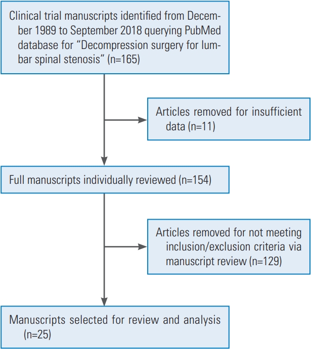
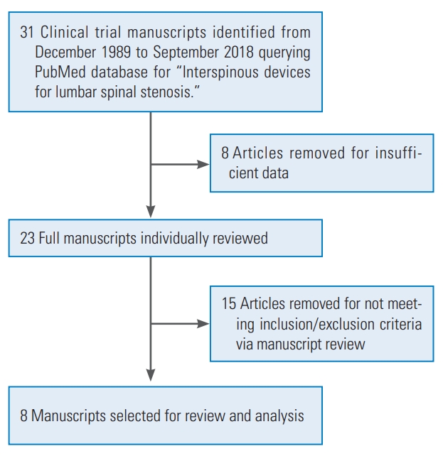
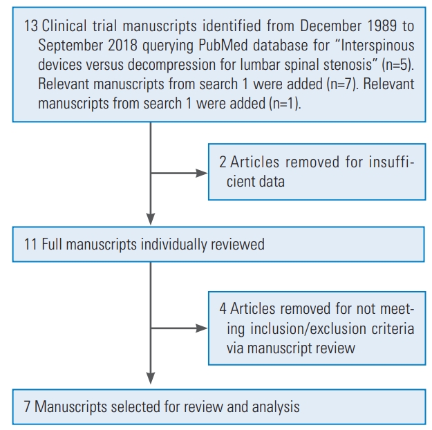
A systematic review was performed to analyze the outcomes from decompression surgery and ISDs using PubMed and included articles dating from December 1989 to August 2018. The search term used for the first part of this review was “Decompression surgery for lumbar spinal stenosis.” The search term used for the second part of this review was “Interspinous devices for lumbar spinal stenosis.” The search term used for the third part of this review was “Interspinous devices versus decompression for lumbar spinal stenosis.” These searches yielded a total of 208 results. Of these, 20 articles were excluded due to insufficient data or inability to access full-text articles in English. Articles were included within this review if they presented primary human data on decompression surgery or ISDs for lumbar spinal stenosis. Only prospective cohort studies, case series, and randomized controlled trials that quantitatively evaluated outcomes using the Visual Analog Scale (VAS), Oswestry Disability Index (ODI), or Japanese Orthopedic Association (JOA) scores were included. Clinical studies that involved other surgical procedures were excluded. According to these criteria, 40 experimental studies met the inclusion criteria.
The extracted clinical articles were then divided into three categories, as follows: (1) studies that described only decompression (Fig. 1), (2) studies that described only ISDs (Fig. 2), and (3) studies that described both ISDs and decompression (Fig. 3). All studies were evaluated for sample size, average age, type of decompression surgery or ISD, functional recovery via VAS, ODI, or JOA score, complications, mortality, and average follow-up or follo-wup range.
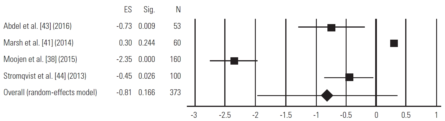
ProMeta 3 was used to generate a random-effects model to analyze the pooled VAS score outcomes obtained by randomized controlled trials that examined differences in preoperative and 1–2-year postoperative VAS pain scores. A positive effect size indicated that ISD surgery was associated with a greater reduction in VAS pain scores compared with lumbar decompression, whereas a negative effect size indicated that lumbar decompression was associated with a greater reduction in pain scores compared with ISD surgery. The longest available postoperative data presented in a given study were used for this analysis. If articles only presented differences between preoperative and postoperative VAS pain scores rather than raw data for postoperative pain scores, the standard deviation for those differences in pain scores was used to model the standard deviation of the postoperative pain scores. A p-value less than 0.05 was considered statistically significant.

Results

1. Studies exclusive to decompression

Twenty-five studies that singularly described the use of decompression surgery for lumbar spinal stenosis were included. In all, these studies involved 3,386 patients, including 1,395 males and 1,895 females, whereas two studies did not report the gender [15,16]. The average participant age was 68.7 years (range, 31–88 years). Types of decompression surgery included Marmot operation [17], minimally invasive [4-12], traditional laminectomy [15-25], unilateral laminectomy [16], open laminectomy [8], endoscopic interlaminar approach [9], endoscopic unilateral decompression [9], lumbar spinous process splitting, midline decompression, mild percutaneous decompression [26], mild interlaminar decompression, windows technique laminoforaminotomy [22], bilateral laminotomy [25], and unilateral laminotomy [16,19,25,27]. The average follow-up period for these studies was 17.6 months (range, 0–90 months). The major findings included decreased neuropathic pain as measured by VAS [4,9,10,15,19-24,26-29], decreased functional impairment secondary to lower back pain as measured by ODI [6-11,16,18,21-23,26,27,29], and reduction in severity of lower back pain as measured by JOA score [4,5,11,19,28] (Table 1). Studies also reported complications associated with decompression such as dural tear [5,8,9,11,12,16,19,21-24] (n=65/3,386, 2.2%), transient neuralgia [5,7,9,11,12] (n=26/3,386, 0.77%), hematoma at the epidural or peridural levels [5,8,9,16,23,25] (n=16/3,386, 0.47%), infection (respiratory, wound, urinary tract) [5,8,11,18,21,22,25] (n=87/3,386, 2.6%), procedural hemorrhage [6,7,18] (n=3/3,386, 0.089%), allergy [18] (n=2/3,386, 0.059%), micturition problems [8,9,20] (n=27/3,386, 0.80%), cardiovascular complications including myocardial infarction, pulmonary emboli, and deep vein thromboses [8,21] (n=5/3,386, 0.15%), foot drop [9] (n=3/3,386, 0.089%), pruritus [20] (n=3/3,386, 0.089%), nausea and vomiting [20] (n=7/3,386, 0.21%), gastric ulcers [21] (n=3/3,386, 0.089%), and degenerative spinal instability [22] (n=6/3,386, 0.18%). Reoperation was required in a few cases [9,18,22,23,25] (n=16/3,386, 0.47%). Only one study reported a perioperative fatality caused by myocardial infarction 1 month after surgery [21].

2. Studies exclusive to interspinous devices

Eight studies that singularly described the use of ISDs for lumbar spinal stenosis were included. In all, these studies involved 1,496 patients, including 807 males and 578 females, whereas two studies did not report the gender [30,31]. The average participant age was 65.1 years (range, 19–89 years). The types of ISDs included percutaneous interspinous process spacer [32] (n=80/1,496, 5.3%), Superion [31,33,34] (n=404/1,496, 27.0%), X-stop [30,33-36] (n=911/1,496, 60.9%), and Aperius (n=156/1,496, 10.4%). The average follow-up time was 23.1 months (range, 1–84 months). The major quantitative findings of ISD treatment for lumbar stenosis included reduction of the VAS score [30,31,33,35,37] and a decrease in the ODI score [30,31,33,34,36] (Table 2). Unlike in the decompression studies, the JOA score was not used to evaluate changes after ISD use. Complications resulting from ISDs were postoperative back pain [34,37] (n=101/1,496, 6.8%), leg pain including sciatica [34,37] (n=80/1,496, 5.3%), spinous process fracture [32,34,35,37] (n=56/1,496, 3.7%), buttock/groin pain [34] (n=22/1,496, 1.5%), cerebrospinal fluid leakage [35] (n=9/1,496, 0.60%), infection including in both superficial skin and deep wounds [33,35] (n=15/1,496, 1.0%), device dislocation [32,35] (n=19/1,496, 1.3%), reoperation [33] (n=18/1,496, 1.2%), removal of the ISD (n=14/1 (n=14/1,496, 0.94%), hematoma (n=1/1,496, 0.07%), and spinal claudication [37] (n=3/1,496, 0.20%).

3. Studies that compared interspinous device and decompression

Seven studies compared the efficacy of decompression versus that of ISD for lumbar spinal stenosis through a randomized controlled trial design or a prospective clinical study. In all, 624 patients were included in these studies: 310 underwent decompression and 314 received an ISD. The average age could not be calculated because some studies included a median age instead of an average age [38,39]. Approaches for decompression included minimally invasive [40], primary lumbosacral [41] spinal bony decompression [38], posterior midline approach [42], and traditional [39,43,44]. The types of ISDs used included Coflex [43], X-stop [40,43,44], Wallis implant [41], interspinous dynamic stabilization devices [42], and a foraminal enlargement lumbar interspinous distraction device [39]. In one study, the type of ISD used was not specified [38]. The average follow-up time in these studies was 18.9 months (range, 0–40 months).
All seven clinical trials demonstrated a statistically significant decrease from the preoperative VAS or ODI scores to the postoperative VAS or ODI scores within each treatment group [38-44]. With regards to improvement in neuropathic pain as measured by the VAS, some studies showed greater improvements between preoperative and the last follow-up after decompression compared with ISD surgery [38,43,44]. With regards to back pain-induced functional abnormalities, one study showed greater improvement between preoperative and the last follow-up after decompression compared with ISD [1]. However, one study showed the opposite effect [40,43] (Table 3). Differences in complication rates between decompression and ISDs were variable in each study. While some studies showed a higher rate of reoperation with ISDs [38,43], others showed the opposite [44] or similar reoperation rates [40]. Dural tears were almost exclusively seen in the decompression group [38-41,43,44], with only a single study reporting one incident of a dural tear in the ISD group [1]. Spinous process fractures were also exclusively seen in the ISD group, with no reported fractures in any of the decompression groups [38-41,43,44] (Table 4).
Several other important factors in the selection of the surgical procedure are the reoperation rates, estimated blood loss, and operative times. Not every clinical trial reported reoperation rates, and for the data that were collected, the results were mixed in terms of the differences in the reoperation rates between decompression surgery and ISD insertion surgery. A 0.47% reoperation rate was found in the studies exclusive to decompression surgery [9,18,22,23,25] and a 1.2% reoperation rate was found in the studies exclusive to ISD surgery [33]. However, studies directly comparing the two treatments reported a 3.9% reoperation rate with decompression surgery and a 8.3% reoperation rate with ISDs [38,40,43,44]. Similarly, estimated blood loss and operation times were inconsistently reported in the clinical trials that were reviewed. The average operation time in decompression-only trials was 92.30±54.78 minutes [8]. The average operation time in ISD-only trials was 43.33±8.50 minutes [37]. In trials that directly compared decompression surgery and ISD surgery, the average operation time was 77.50±41.44 minutes for the former procedure [37] and 57.55±45.44 minutes for the latter procedure [37]. The average intraoperative blood loss in decompression-only studies was 110.18±88.74 mL [38]. The average intraoperative blood loss in ISD-only studies was 23.33±17.56 mL [37]. In trials that directly compared decompression surgery and ISD surgery, the average intraoperative blood loss for the former procedure was 262±0 mL [37] and 143.07±125.96 mL [37] for the latter procedure. Two studies provided categorized data rather than numerical data for blood loss, but in both studies, a characteristic of decompression trials was greater blood loss compared with ISD surgery [37].

4. Meta-analysis

Four randomized controlled trials assessed the differences in the preoperative and 1–2-year postoperative VAS scores between ISD surgery and lumbar decompression [38-41,43,44]. According to a random-effects model, the difference between the preoperative and pooled 1–2-year postoperative VAS scores between ISD surgery and lumbar decompression was not statistically significant (p=0.166). The results from the random-effects model are graphically displayed as a forest plot in Fig. 4.

Discussion

This comprehensive systematic review investigated three groups of studies that examined the clinical and functional impact of decompression surgery or placement of ISDs for lumbar spinal stenosis. Of the literature that explored the role of decompression exclusively, most studies compiled within this review involved conventional decompression surgery via open laminectomy [8,15,16,18-24,27] or laminotomy [5,9,11,25], but other types of decompression surgery were explored, including minimally invasive decompression [4,6-8], full-endoscopic interlaminar bilateral decompression [9], the Marmot operation [17], lumbar spinous process splitting decompression, midline Author decompression, modified unilateral laminotomy for bilateral decompression, percutaneous minimally invasive lumbar decompression [26], “Windows” decompression [22], and ultra-minimally invasive lumbar decompression [10]. Minimally invasive decompression surgery is associated with comparable decreases in ODI scores as is open laminectomy, but this procedure caused fewer surgical complications including dural tears, postoperative hematomas, wound infections, and UTIs according to these studies [8]. Generally, regardless of the decompressive surgical approach, studies consistently demonstrated improvement in VAS, ODI, and JOA scores. Infection was the most common complication in clinical studies that exclusively examined decompression surgery [5,8,9,18,21].
Papers that reviewed the use of ISDs for the surgical treatment of lumbar spinal stenosis mainly used the following types: the Superion, X-stop, percutaneous interspinous process spacers, and Aperius devices. Of these, X-stop was reviewed the most (five clinical studies) [30,33-36], while Superion, ISPs, and Aperius were reviewed in three [31,33,34], one [32], and one [37], respectively. Both the Superion and X-stop devices showed comparable decreases in ODI and VAS scores, and varying similarities and differences were observed in postoperative complications. In several studies, X-stop cases were associated with a higher percentage of back and leg pain and wound infections but fewer incidences of spinous process fractures [34]. Of the two, the Superion device allows for a more minimally invasive approach and thus reduced disruption of neighboring spinal structures, which may account for the reduced postoperative pain observed in these patients [34]. Additionally, the Superion device has thicker wings than the X-stop device, which may increase long-term stability and reduce incidences of repeat surgery [34]. Across all studies that directly compared the two ISD devices, the Superion and X-stop implants had similar clinical outcomes, which suggests that preference for one surgery over the other might be indicated by surgeon familiarity and preference [33,34]. Complications and patient complaints necessitated the removal of nearly 10% of all Aperius implants in one clinical study. The most common complication within ISD-exclusive surgeries was spinous process fracture, which may be improved with increased surgeon familiarity and experience with placing the ISD [32,34-37].
In the seven studies that directly compared decompression surgery with ISD insertion, no clear consensus as to the superiority of either surgical intervention was reported. All studies showed statistically significant improvement in postoperative VAS and ODI scores within both treatment arms [38-44]. Across all six studies, the VAS and ODI scores were also comparable between the two groups; neither decompression nor ISD surgery consistently demonstrated superiority in either VAS or ODI scores. Three clinical studies demonstrated a greater change in VAS or ODI scores for decompression patients [38,43,44], two clinical trials demonstrated a greater change in VAS scores for ISD patients [39,40], and two clinical trials demonstrated no significant difference in the changes in VAS or ODI scores for decompression versus ISD patients [41,42]. One study reported 17 more repeat surgeries in the group that received ISD device insertion surgery than in the group that received decompression surgery, even with nearly equal patient sizes [38]. However, another study reported fewer incidences of repeat surgery and postoperative complications in the X-stop group than in the decompression group. Some discrepancies were reported in the complications among studies. One study with a patient size of 60 mentioned no major complications, while other studies mentioned unique complications such as postoperative cauda equina syndrome and foot drop, which did not occur as a result of any other similar surgeries [40,41,43]. Similarly, the most common complication within clinical trials that examined decompression and ISD device insertion was reoperation [38,40,43,44]. A higher reoperation rate was observed in the ISD group compared with the decompression group, which suggests that decompression surgery has more favorable long-term results, at least in terms of this metric. Similarly, the ISD-exclusive studies reported a higher reoperation rate than the decompression surgery-exclusive studies. The heterogeneity in outcomes data between ISDs and lumbar decompression was reflected within our random-effects model. While overall postoperative reductions in VAS scores were identified between ISDs and lumbar decompression, our model did not find a statistical difference between ISD surgery and lumbar decompression to cause a greater reduction in VAS scores compared with the other intervention.
Operation times and intraoperative blood loss can also be compared. The average operation time was consistently lower for ISD surgery than for decompression surgery, which may be beneficial for patients who cannot tolerate long operation times [8]. This conclusion was confirmed by the clinical trials that directly compared decompression surgery and ISD surgery. In all cases, these clinical trials reported longer operation times for decompression surgery [37]. Similarly, every clinical trial that directly compared the two treatment mechanisms reported less intraoperative bleeding for ISDs than for decompression surgery [37]. Incidentally, one clinical trial reported no measurable blood loss for decompression surgery primarily because there was continuous irrigation during surgery and thus no drainage was required [8,45]. Therefore, ISD surgery may be recommended for patients who benefit from shorter operation times and/or who are at greater risk for blood loss complications.
A concern with the use of ISDs is their potential to introduce segmental kyphosis through an increase in interspinous flexion and distraction as the mechanism of indirect decompression. This has led to significant concerns on how placement of ISDs can potentially negatively affect overall sagittal balance. Several biomechanical studies have been performed to investigate segmental tilt after implantation of an ISD [46-48]. Wilke et al. [47] found that while the DIAM device introduced kyphosis into the operated segment, the Coflex, Wallis, and X-stop devices had no effect on the 0.5°–0.7° of kyphosis. Another biomechanical study by Anasetti et al. [48] reported similar findings, as the DIAM device induced a shift toward kyphosis at the implanted level. Alfieri et al. [49] performed a systematic review of clinical studies that involved ISD and described the risk of introducing kyphosis at the operated lumbar segment. They identified 11 studies where kyphosis was measured preoperatively and postoperatively and reported no differences in the segmental angle. They concluded that no clinical data suggested that ISD significantly introduces kyphosis. Schulte et al. [50] has performed the only prospective study that evaluated sagittal balance in 20 patients who received an X-stop device. Using postoperative 91.44-cm films, they found an average improvement in the overall sagittal alignment of −2.0cm. Their hypothesis was that a postoperative relief of stenosis was achieved, which allowed the spine to assume the most efficient posture and sagittal balance through the entirety of the lumbar spine, with only a segmental reduction in extension at the operated level. Although ISDs have a potential locally segmental kyphotic effect, there appears to be the potential for improvement in neurogenic posture from relief of claudication symptoms that may maintain overall global sagittal alignment.
In this review, we attempt to distill studies on decompression surgery or placement of an ISD for lumbar spinal stenosis as they relate to postoperative outcome and complication rates. The theoretical benefit of an ISD over traditional decompression lies in the potential for a more minimally invasive approach with a motion-preserving implant to relieve the symptoms of neurogenic claudication. ISDs may also be helpful in patients who cannot tolerate lengthy procedures under general anesthesia due to medical comorbidities. ISD surgery was consistently associated with shorter operation times and less intraoperative blood loss compared with decompression surgery, both in decompression-only and interspinous-only papers and in papers that assessed both treatment types. For prospective, technique-exclusive, or comparison studies, we found that ISD surgery leads to favorable postoperative functional outcomes in terms of VAS and ODI scores but that it does not demonstrate superiority over decompression surgery. Incidences of 5.3%, 1.3%, and 8.3% in postoperative leg pain, device dislocation, and reoperation rate, respectively, were observed. Overall, we recommend the use of decompression surgery based on the lower reoperation rates. Lower reoperation rates were consistently reported in the decompression surgery-exclusive papers and the papers that compared ISD versus decompression surgery.
Limitations of this study include inconsistent reporting of measurements among studies. Inconsistencies were also found in the extent of complications reported, with more exhaustive studies reporting unique complications, while some studies simply stated that no major complications were encountered [10,15,26,29,30,41,42]. Another limitation of this paper is the variation in postoperative care, which is important for long-term complications such as reoperation rates. Despite these limitations, we believe the information provided in this review puts into perspective the overall functional outcomes and reported complication rates in prospective studies of decompression versus ISD surgery for lumbar spinal stenosis. As with all clinical treatments, careful patient selection with an appropriately tailored surgical intervention remains crucial for successful patient outcomes.

Conclusions

Treatment of lumbar spinal stenosis can be accomplished through decompression surgery or through the insertion of an ISD. Although studies reported higher rates of dural tears in decompression surgery and higher reoperation rates after ISD surgery, a meta-analysis revealed no difference in functional outcome, as improvements were seen in both groups. Careful patient selection remains crucial for either surgical procedure to ensure optimal surgical outcomes tailored to each patient. More diverse studies are needed to determine the superiority of one technique over the other for different patient populations.

Conflict of Interest

No potential conflict of interest relevant to this article was reported.

Fig. 1.
Flowchart for screening manuscripts that reported “Decompression surgery for lumbar spinal stenosis.” In all, 165 clinical studies were initially identified from the PubMed database. After the inclusion and exclusion criteria were applied, 25 manuscripts (11 randomized controlled trials, 11 prospective cohort studies, and three case series) were selected for review and analysis.
asj-2019-0105f1.jpg
Fig. 2.
Flowchart for screening manuscripts that reported “Interspinous devices for lumbar spinal stenosis.” Thirty-one clinical studies that investigated the role of interspinous device for lumbar spinal stenosis were initially identified in the PubMed database. After the inclusion and exclusion criteria were applied, eight manuscripts (four randomized controlled trials, three prospective cohort studies, and one case series) were selected for review and analysis.
asj-2019-0105f2.jpg
Fig. 3.
Flowchart for screening manuscripts that reported “Interspinous devices versus decompression for lumbar spinal stenosis.” Twelve clinical studies that compared interspinous device with decompression for lumbar spinal stenosis were initially identifi ed in the PubMed da- umbar spinal stenosis were initially identifi ed in the PubMed da- pinal stenosis were initially identifi ed in the PubMed da- pinal stenosis were initially identifi ed in the PubMed da- tenosis were initially identifi ed in the PubMed da- tenosis were initially identifi ed in the PubMed da- were initially identified in the PubMed database, and eight relevant studies from previous queries were added. After the inclusion and exclusion criteria were applied, seven manuscripts (one prospective cohort study and six randomized controlled clinical trials) were selected for review and analysis.
asj-2019-0105f3.jpg
Fig. 4.
Forest plot depicting the random-effects model of the differences in preoperative and postoperative VAS scores between ISD use and lumbar decompression. After pooling outcomes from the four randomized trials with VAS outcomes data, a statistically significant difference between the preoperative and 1–2-year postoperative VAS scores between ISD and lumbar decompression was not identified (p=0.166). A positive effect size indicates a greater reduction in VAS scores in the ISD group compared with the lumbar decompression group, while a negative effect size indicates a greater reduction in VAS scores in the lumbar decompression group compared with the ISD group. VAS, Visual Analog Scale; ISD, interspinous device; ES, effect size; Sig, statistical significance.
asj-2019-0105f4.jpg
Table 1.
Clinical outcomes after decompression surgery for lumbar spinal stenosis (N=25 clinical trials)
Author Year Type of clinical trial Type of decompressive surgery Sample size Age (yr) VAS ODI JOA Mortality/complications FU
Minamide et al. [5] 2018 PCS Microendoscopic laminotomy 242 (101 M, 141 F) Average, 68.1; range, 46 to 85 NA NA Preop, 14.4±4.3; postop at last FU, 23.3±4.8 Dural tear (3); transient neuralgia (3); epidural hematoma (4); infection (1) Average: 4.6 yr
Minamide et al. [4] 2017 PCS MID 122 (57 M, 65 F) Average, 70.4±8; range, 46 to 88 Preop, 55.9±28.1; postop at last FU, 35.0±30.1 NA Preop, 13.4±4.4; postop at last FU, 20.5±5.3 NA Average: 2.4 yr
Benyamin et al. [6] 2016 RCT MID 149 (74 M, 75 F) Average, 75.6±7.0 NA Preop, 53.0±12.9; postop at 1 yr, 37.0 NA Non-serious procedural hemorrhage (1) 6 mo, 1 yr, 2 yr
Staats et al. [7] 2016 RCT MID 149 (74 M, 75 F) Average, 75.6±7.0 NA Preop, 53.0±12.9; postop at 6 mo, 34.5 NA Procedural hemorrhage (1); procedural pain (1) 6 mo
Delitto et al. [18] 2015 RCT Traditional decompression with laminectomies, facet resection and neuroforaminotomy 87 (44 M, 43 F) Average, 66.6±10.5 NA Preop, 42.6; postop at 24 mo, 25.2 NA Allergy (1); blood loss (1); respiratory tract infection (3); reoperation of lumbar spine (3); worsening symptoms (2); wound healing/site infection (7) 24 mo
Nerland et al. [8] 2015 PCS MID, open laminectomy MID: 471 (249 M, 222 F); laminectomy: 414 (209 M, 205 F) MID: average, 66.6; laminectomy: average, 70.1 NA MID: preop, 38.0; postop at 1 yr, 19.6; laminectomy: preop, 42.1; postop at 1 yr, 25.0 NA MID: dural tear/spinal fluid leak (11); postop hematoma (1); cardiovascular complications (1); wound infection (8); UTIs (17); micturition problems (3); pneumonia (5); Laminectomy: dural tear/spinal fluid leak (21); postop hematoma (4); anaphylactic reaction (1); wound infection (11); UTIs (20); micturition problems (15); pneumonia (2); pulmonary embolism (1); DVT (2) 10 wk, 6 mo, 12 mo, 24 mo
Komp et al. [9] 2015 RCT MI, FI Total: 180 (91 M, 69 F); MI: 80; FI: 80 Total: average, 62; range, 41 to 84 MI: preop, 25; postop at 24 mo, 12; FI: preop, 23; postop at 24 mo, 17 MI: preop, 88; postop at 24 mo, 30; FI: preop, 84; postop at 24 mo, 28 NA MI: transient dysesthesia (7); transient urinary retention (3); dura injuries (3); increased foot dorsiflexion paresis (2); epidural hematoma (1); delayed wound healing (2); soft tissue infections (2); revision surgery with fusion (2); FI: transient dysesthesia (4); transient urinary retention (1); dura injuries (2); increased foot dorsiflexion paresis (1); delayed wound healing (1); revision surgery (1) 3 mo, 6 mo, 12 mo, 24 mo
Mobbs et al.[16] 2014 RCT ULBD, traditional laminectomy ULBD: 27; laminectomy: 27 ULBD: average, 72.7±10.4; laminectomy: average, 65.8±14.3 NA ULBD: preop, 51.4±19.4; mean improvement at last FU, 28.6±27.7; laminectomy: preop, 46.6±18.9; mean improvement at last FU, 17.8±15.4 NA ULBD: dural tear (1); laminectomy: foot drop (1); hematoma (1); dural tear (1) ULBD: average: 36.9±4.3 mo; laminectomy: average: 44.3±15.0 mo
Hajasek-aran et al. [28] 2013 HCT LSPD, midline decompression LSPD: 28 (16 M,12F); midline decompression: 23 (14M,9F) LSPD: average, 57.25±11.23; range: 35 to 76; midline decompression: average, 54.48±8.21; range, 39 to 71 LSPD: preop, 5.35; postop at last FU, 2.46; midline decompression: preop, 5.43; postop at last FU, 2.96 NA LSPD: preop, 6.96; postop at last FU, 10.75; midline decompression: preop, 6.69; postop at last FU, 11.39 No major complications Average: 14.2±2.9 mo
Liu et al. [19] 2013 RCT M-ULBD, traditional laminectomy M-ULBD: 27 (15 M, 12 F); laminectomy: 29 (18 M, 11 F) M-ULBD: average, 59.4±4.7; range, 49 to 71; laminectomy: average, 61.1±3.1; range, 52 to 69 M-ULBD: preop, 5.6±1.7; postop, 1.0±0.5; laminectomy: preop, 7.9±1.3; postop at last FU, 2.6±0.7 NA M-ULBD: preop, 11.6±2.6; postop, 26.7±2.1; laminectomy: preop, 10.9±1.0; postop at last FU, 24.3±2.5 M-ULBD: dural tear (3); laminectomy: dural tear (1) Average: 2yr
Deer et al, [26] 2012 PCS Mild percutaneous lumbar decompression 46 (17 M, 29 F) Average, 66.1; range, 46 to 80 Preop, 6.9±0.6; postop at 1 yr, 4.0±1.0 Preop, 49.4±2.5; postop at 1 yr, 32.0±5.8 NA No major complications 12 wk, 6 mo and 1 yr
Guilfoyle et al. [20] 2012 RCT Traditional laminectomy w/o epidural fentanyl, traditional laminectomy with epidural fentanyl Decompression w/o fentanyl: 31 (14 M,17 F); decompression with fentanyl: 29 (16 M,13 F) Decompression w/o fentanyl: average, 65.7±11.5; decompression with fentanyl: average, 69.9±9.8 Decompression w/o fentanyl: preop, 4.1±3.0; postop in recovery room, 4.7±2.4; decompression with fentanyl: preop, 3.4±2.9; postop in recovery room, 2.6±2.7 NA NA Decompression w/o fentanyl: itching (2); nausea/vomiting (4); decompression with fentanyl: itching (1); nausea/vomiting (3); urinary catheter (3) In “recovery”, before postop day 1
Wong et al.[29] 2012 Case series Mild interlaminar decompression 17 (10 M,7 F) Average, 73.1; range, 63 to 86 Preop, 7.6; postop at 1 yr, 2.3 Preop, 48.4; postop at 1yr, 21.7 NA No major complications 1 yr
Komp et al. [45] 2011 Case series Full-endoscopic unilateral decompression 90 (41 M, 49 F) Average, 61; range, 43 to 81 Preop, 23; postop at 24 mo, 17 Preop, 85; postop at 24 mo, 31 NA Dysesthesia (5); urinary retention (2); dural injuries (2); increase in foot dorsiflexion paresis (1); repeat surgery (2) 3 mo, 6 mo, 12 mo, 24 mo
Jakola et al.[21] 2010 PCS Traditional laminectomy 101 (51 M, 50 F) Average, 75.3±4.1; range, 70 to 86 Preop, 55.7; postop at 12 mo, 35.9 Preop, 44.2; postop at 12 mo, 27.9 NA Dural tear (9); superficial wound infection (3); perioperative death (1); UTI (1); deep wound infection (2); myocardial infarction (1); gastric ulcer (3) 3 mo, 12 mo
Chopko et al.[10] 2010 PCS Ultra-minimally invasive lumbar decompression 75 (29 M, 46 F) Average, 70.0; range, 37 to 88 Preop, 7.3; postop at 6 wk, 3.7 Preop, 47.4; postop, 29.5 NA No major complications 6 wk
Pao et al. [11] 2009 PCS Microscopic decompressive laminoto-my 53 (17 M, 36 F) Average, 62.0; range, 36 to 86 NA Preop, 64.3±20.0; postop at last FU, 16.7±20.0 Preop, 9.4±6.1; postop at last FU, 24.2±6.0 UTI (2); dural tear (5); wrong level operation (2); transient neuralgia (4) Average: 15.7 mo
Fu et al. [22] 2008 PCS “Windows technique” laminoforaminotomy, traditional laminectomy Windows: 76 (37 M, 39 F); laminectomy: 76 (33 M, 43 F) Windows: average, 57; range, 47 to 70; laminectomy: average, 57; range, 45 to 73 Windows: preop, 1.14±0.96; postop at last FU, 0.05±0.22; laminectomy: preop, 1.17±0.91; postop at last FU, 0.63±1.07 Windows: preop, 39.26±6.54; postop at last FU, 0.37±0.96; laminectomy: preop, 39.76±6.94; postop at last FU, 3.37±8.55 NA Windows: dural tear (3); UTI (2); laminectomy: dural tear (2); UTI (2); degenerative spinal instability (6); repeat surgery with fusion (4) Average: 40 mo
Ca vuşoğlu et al. [27] 2007 PCS Unilateral laminectomy 50 (21 M, 29 F) Average, 69.81±15.15; range, 43 to 82 Preop, 6.92±1.04; postop at last FU, 2.16±0.81 Preop, 31.14±9.27; postop at last FU, 14.02±9.27 NA None Average: 22.8 mo
Cho et al. [17] 2007 RCT Marmot operation, traditional laminectomy Marmot: 40 (16 M, 24 F); laminectomy: 30 (15 M, 15 F) Marmot: average, 61.2±10.8; range: 52 to 77; laminectomy: average, 58.8±14.9; range, 42 to 70 Marmot: preop, 6.45±2.60; postop, 2.38±1.89; laminectomy: preop, 7.07±1.49; postop, 4.00±2.00 NA Marmot: preop, 7.10±2.84; postop, 13.0±1.97; laminectomy: preop, 8.44±2.59; postop, 11.4±3.20 No major complications Marmot: average: 15.1 mo; laminectomy: average: 14.8 mo
Malmivaara et al. [23] 2007 RCT Traditional laminectomy 50 (11 M,39F) Average, 63±9 Preop, 6.7; postop at 2 yr, 2.9 Preop, 33,0; postop at 2yr, 22.8 NA Dural tear (7); misplaced transpedicular screw (1); peridural hematoma (1); respiratory distress (1); repeat surgery (1) 6 mo, 1 yr, 2 yr
Thome et al. [25] 2005 RCT Bilateral laminotomy (group 1); unilateral laminotomy (group 2); laminectomy (group 3) G1 : 40 (20 M, 20 F); G2: 40 (15 M, 25 F); G3: 40 (18 M, 22 F) G1: average, 70±7; G2: average, 67±6; G3: average, 69±10 G1 : preop, 7.5±2.3; postop at final FU, 2.3±2.4; G2: preop, 7.5±2.3; postop at final FU, 3.6±2.7; G3: preop, 7.5±2.3; postop at final FU, 4±1 NA NA G1: incidental durotomy (2); G2: incidental durotomy (5); increased radicular deficit (2); repeat surgery (2); epidural hematoma (2); G3: incidental durotomy (8) repeat surgery (2); epidural hematoma (2); wound infection (1) 3 mo, 6 mo, 12 mo
Ikuta et al, [12] 2005 Case series Microendoscopic posterior decompression 47 (23 M,24 F) Average, 66; range, 31 to 88 NA NA Preop, 14.3±4.1; postop at final FU, 24.9±3.2 Du ral tear (4); intra-articular fracture (3); neurological deficit (7) Average: 22 mo
Konno et al. [15] 2000 PCS Traditional laminectomy 42 Average, 63 Preop, 7.4; postop at 3 yr, 3.5 NA NA No major complications 1yr,3yr
Weiner et al, [24] 1999 PCS Traditional laminectomy 50 (29 M,21 F) Average, 72; range, 49 to 88 Preop, 8; postop, 2 NA NA Dural tear (4) 9 mo

Patients were evaluated for change in preop and postop VAS, ODI, and JOA scores and postoperative complications.

VAS, Visual Analog Scale; ODI, Oswestry Disability Index; JOA, Japanese Orthopedic Association; FU, follow-up; PCS, prospective cohort study; M, male; F, female; NA, data was not reported for that category; Preop, preoperative; Postop, postoperative; MID, minimally invasive decompression; RCT, randomized controlled clinical trial; UTI, urinary tract infection; DVT, deep vein thrombosis; MI, microsurgical laminotomy; FI, full-endoscopic interlaminar bilateral decompression; ULBD, unilateral laminectomy for bilateral decompression; LSPD, lumbar spinous process splitting decompression; M-ULBD, modified unilateral laminotomy for bilateral decompression; w/o, without.

Table 2.
Clinical outcomes after interspinous device use for lumbar spinal stenosis (N=8 clinical trials)
Author Year Type of clinical trial Type of interspinous device Sample size Age (yr) VAS ODI Mortality/complications FU
Nunley et al. [31] 2017 HCT Superion 89 NA Preop to postop % VAS leg change: 73% Preop to postop % change: 73% NA 4 yr
Marcia et al. [32] 2015 Prospective cohort study Percutaneous interspinous process spacer 80 (39 M, 41 F) Average, 70.4±8.7; range, 51 to 85 Preop, 8.1±2; postop at 1 mo, 4.4±2 Preop, 23.3±10; postop at 1 mo, 11.7±8.5 Dislocation (1); spinous process fracture (2) 1 mo, 1 yr, 3 yr
Patel et al, [34] 2015 HCT Superion, X-stop Superion: 190 (110 M, 80 F); X-stop: 201 (129 M,72 F) Superion: average, 67±9; range, 47 to 88; X-stop: average, 66±10; range, 46 to 89 NA Superion: preop, 39±13; postop, 20±18; X-stop: preop, 40±12; postop, 18±15 Superion (after 2 yr): back pain (49); leg pain (33); spinous process fracture (23); buttock/groin pain (10); X-stop (after 2 yr): back pain (61); leg pain (45); spinous process fracture (13); buttock/groin pain (12) 6 wk, 3 mo, 6 mo, 12 mo, 18 mo, 24 mo
Puzzilli et al. [35] 2014 RCT X-stop Total: 542 (289 M, 253 F); X-stop: 422; non-surgical conservative treatment: 120 Total: average, 63.6; range, 45 to 82 X-stop: preop, 8.3; postop at 7 yr, 2.7 NA Total: spinous process fracture (16); cerebrospinal fluid leakages (9); superficial skin infection (13); device dislocation (18) 1 mo, 6 mo, 1 yr, 2 yr, 3 yr, 4 yr, 5 yr, 6 yr, 7 yr
Patel et al, [33] 2014 HCT Superion, X-stop Total: 192 (150 M, 42 F); superion: 101; X-stop: 91 Total: average, 67 Superion: preop, 59±26; postop, 21±26; X-stop: preop, 55±26; postop, 21±25 Superion: preop, 37±12; postop, 18±16; X-stop: preop, 39±12; postop, 20±16 Superion: repeat surgery (18); X-stop: deep wound infection (2); repeat surgery (18) 6 wk, 3 mo, 6 mo, 12 mo, 18 mo, 24 mo
Ploumis et al. [30] 2012 Case series X-stop 22 Average, 64.5; range, 57 to 71 Preop, 4.5±1.0; postop at last FU, 2.5±0.7 Preop, 50.7±4.4; postop at last FU, 19.9±3.7 No major complications 2 yr
Van Meirhaeghe et al. [37] 2012 Prospective observational study Aperius 156 (78 M, 78 F) Average, 64.8±11.9; range, 19 to 84 Preop, 5.9±2.4; postop at 6 wk, 3.7±2.8 NA Removal of interspinous process decompression (14); spinous process fracture (1); back pain (1); hematoma (1); pain in extremity (1); spinal claudication (3); sciatica (1); arthralgia (6); lumbar spinal stenosis (3) 6 wk, 12 mo
Siddiqui et al. [36] 2007 Prospective observational study X-stop 24 (12 M, 12 F) Median, 71.5 NA Preop, 48; postop at 12 mo, 37 Spinous process fracture (2) 12 mo

Patients were evaluated for change in preop and postop VAS and ODI scores and postop complications.

VAS, Visual Analog Scale; ODI, Oswestry Disability Index; FU, follow-up; RCT, randomized controlled clinical trial; NA, data was not reported for that category; Preop, preoperative; Postop, postoperative.

Table 3.
Comparison of outcomes with decompression surgery versus interspinous device in lumbar spinal stenosis (N=7 clinical trials)
Author Year Type of clinical trial Type of decompression surgery and interspinous device Sample size Age (yr) VAS ODI Mortality/complications FU
Abdel Ghany et al.[43] 2016 Prospective clinical study Traditional laminectomy, Coflex (21), X-stop (7) Laminectomy: 25 (10 M, 15 F); ISP: 28 (22 M, 6 F) Laminectomy: average, 51.8±8.2; ISP: average, 55±10.7 Laminectomy: preop, 4.00±1.00; postop at last FU, 2.96±1.46; ISP: preop, 3.68±1.06; postop at last FU, 3.59±1.39 Laminectomy: preop, 56.48±5.21; postop at last FU, 26.96±12.89; ISP: preop, 54.43±6.49; postop at last FU, 30.67±13.28 Laminectomy: postop instability (3); dural tear (3); wrong level (1); meningitis (1); ISP: repeat surgery (6); spinous erosion (6); rotated X-stop (3); dural tear (1); wound infection (1); foot drop (1); wrong level (1) 12 mo
Mohar et al.[42] 2016 RCT Decompression: (posterior midline approach, flavectomy and laminotomy with possible additional partial facetectomy and/or foraminotomy, IDSDs Decompression: 6 (2 M, 4 F); IDSD: 6 (4 M, 2 F) Decompression: average, 51; range, 39 to 61; IDSD: average, 49; range, 36 to 55 No statistically significant differences in pre- and postop ODI, VAS-lumbar and VAS-leg scores between groups No statistically significant differences in pre- and postop ODI, VAS-lumbar and VAS-leg scores between groups No major complications Decompression: 188 day; IDSD: 196 day
Moojen et al. [38] 2015 RCT Spinal bony decompression, unnamed ISP Decompression: 79 (37 M, 42 F); ISP: 80 (49 M, 31 F) Decompression: median, 64; range, 47 to 83; ISP: median, 66; range, 45 to 83 Decompression: preop, 52; postop at 104 wk, 28; ISP: preop: 50; postop at 104 wk, 36 NA Decompression: epidural hematoma (2); dural tear (4); repeat surgery (6); ISP: spinous process fractures (3); wrong level (1); repeat surgery (23) 2,4, 8,12, 26, 52, 104 wk
Lome et al.[40] 2015 RCT MID, X-stop MID: 41 (23 M, 18 F); X-stop: 40 (17 M, 23 F) MID: average, 67±8.7; X-stop: average, 67±8.8 NA MID: preop, 33.8±2.5; postop at 2 yr, 18.4±2.6; X-stop: preop, 32.9±2.7; postop at 2 yr, 14.3±2.7 MID: hematoma (3); postop cauda equine syndrome (1); dural tear (2); urine retention (1); repeat surgery (3); X-stop: spinous process fracture (1); late fracture (1); dislocation of X-stop (1); repeat surgery (3) 6 wk, 3 mo, 1 yr, 2 yr
Marsh et al.[41] 2014 RCT Priman/lumbosacral decompression,Wallis implant Decompression: 30 (14 M, 16 F); Wallis: 30 (16 F, 14 M) Decompression: average, 56.4±12.9; range, 34 to 76; Wallis: average, 59.6±13.4; range, 35 to 81 Decompression: change from preop to last FU, 2.7±2.4; Wallis: change from preop to last FU, 3.5±3.4 Decompression: change from preop to last FU, 10.6±19.3; Wallis: change from preop to last FU, 19.3±24.0 No major complications Average: 40 mo
Moojen et al.[39] 2013 RCT Traditional decompression, FELIX Decompression: 79 (37 M, 42 F); FELIX: 80 (49 M, 31 F) Decompression: median, 66; range, 45 to 83; FELIX: median, 64; range, 47 to 83 Decompression: preop, 49; postop at 52 wk, 31; FELIX: preop, 60; postop at 52 wk, 23 NA Decompression: epidural hematoma (2); dural tear (4); FELIX: visual disturbance (1); pseudoradicular leg pain (1); interspinous process fractures (3) 2,4, 8,12, 26, 52 wk
Strömqvist et al. [44] 2013 RCT Traditional decompression, X-stop Decompression: 50 (26 M, 24 F); X-stop: 50 (30 M, 20 F) Decompression: average, 71; range, 57 to 84; X-stop: average, 67; range, 49 to 89 Decompression: preop, 60±26; postop at 24 mo, 23±29; Xstop: preop, 59±28; postop at 24 mo, 34±32 NA Decompression: dural injury (3); repeat surgery (3); X-stop: spinous process fracture (1) 24 mo

Patients were evaluated for change in preop and postop VAS and ODI scores and postop complications.

VAS, Visual Analog Scale; ODI, Oswestry Disability Index; FU, follow-up; M, male; F, female; ISP, interspinous process; Preop, preoperative; Postop, postoperative; RCT, randomized controlled clinical trial; IDSD, interspinous dynamic stabilization device; NA, data was not reported for that category; MID, minimally invasive decompression; FELIX, foraminal enlargement lumbar interspinous distraXion.

Table 4.
Complications associated with decompression-exclusive surgery, ISP-exclusive surgery, and ISP versus decompression surgeries
Complication Decompression surgery exclusive studies ISP device exclusive studies ISP vs. decompression surgeries
Decompression ISP device
Dural tear 65/3,386 (2.2) 16/310 (5.2) 1/314 (0.32)
Transient neuralgia 26/3,386 (0.77)
Hematoma at epidural or peridural level 16/3,386 (0.47) 1/1,496 (0.07) 7/310 (2.3)
Infection (respiratory, wound, or urinary tract infection) 87/3,386 (2.6) 15/1,496 (1.0) 1/314 (0.32)
Procedural hemorrhage 3/3,386 (0.089)
Allergy 2/3,386 (0.059)
Micturition problem 27/3,386 (0.80)
Cardiovascular complication (myocardial infarction, pulmonary emboli, deep vein thrombosis) 5/3,386 (0.15)
Increased foot dorsiflexion 3/3,386 (0.089)
Pruritus 3/3,386 (0.089)
Nausea and vomiting 7/3,386 (0.21)
Gastric ulcers 3/3,386 (0.089)
Degenerative spinal instability 6/3,386 (0.18)
Wrong level of operation or misplaced screw 3/3,386 (0.089) 1/310 (0.32) 3/314 (0.10)
Reoperation 16/3,386 (0.47) 18/1,496 (1.2) 12/310 (3.9) 26/314 (8.3)
Back pain 101/1,496 (6.8)
Buttock/groin pain 22/1,496 (1.5)
Spinal claudication 3/1,496 (0.20)
Leg pain 80/1,496 (5.3) 1/314 (0.32)
Spinous process fracture 56/1,496 (3.7) 9/314 (2.9)
Cerebrospinal fluid leakage 9/1,496 (0.60)
Device dislocation 19/1,496 (1.3) 1/314 (0.32)

Values are presented as number/total number (%).

ISP, interspinous process.

References

1. Hall S, Bartleson JD, Onofrio BM, Baker HL Jr, Okazaki H, O’Duffy JD. Lumbar spinal stenosis: clinical features, diagnostic procedures, and results of surgical treatment in 68 patients. Ann Intern Med 1985 103:271–5.
crossref pmid
2. Binder DK, Schmidt MH, Weinstein PR. Lumbar spinal stenosis. Semin Neurol 2002 22:157–66.
crossref pmid pdf
3. Atlas SJ, Delitto A. Spinal stenosis: surgical versus nonsurgical treatment. Clin Orthop Relat Res 2006 443:198–207.
crossref pmid
4. Minamide A, Yoshida M, Iwahashi H, et al. Minimally invasive decompression surgery for lumbar spinal stenosis with degenerative scoliosis: predictive factors of radiographic and clinical outcomes. J Orthop Sci 2017 22:377–83.
crossref pmid
5. Minamide A, Yoshida M, Simpson AK, et al. Minimally invasive spinal decompression for degenerative lumbar spondylolisthesis and stenosis maintains stability and may avoid the need for fusion. Bone Joint J 2018 100-B:499–506.
crossref pmid
6. Benyamin RM, Staats PS, MiDAS Encore I. MILD(R) is an effective treatment for lumbar spinal stenosis with neurogenic claudication: MiDAS ENCORE randomized controlled trial. Pain Physician 2016 19:229–42.
pmid
7. Staats PS, Benyamin RM RM, MiDAS ENCORE Investigators. MiDAS ENCORE: randomized controlled clinical trial report of 6-month results. Pain Physician 2016 19:25–38.
crossref pmid
8. Nerland US, Jakola AS, Solheim O, et al. Minimally invasive decompression versus open laminectomy for central stenosis of the lumbar spine: pragmatic comparative effectiveness study. BMJ 2015 350:h1603.
crossref pmid pmc
9. Komp M, Hahn P, Oezdemir S, et al. Bilateral spinal decompression of lumbar central stenosis with the full-endoscopic interlaminar versus microsurgical laminotomy technique: a prospective, randomized, controlled study. Pain Physician 2015 18:61–70.
crossref pmid
10. Chopko B, Caraway DL. MiDAS I (mild decompression alternative to open surgery): a preliminary report of a prospective, multi-center clinical study. Pain Physician 2010 13:369–78.
crossref pmid
11. Pao JL, Chen WC, Chen PQ. Clinical outcomes of microendoscopic decompressive laminotomy for degenerative lumbar spinal stenosis. Eur Spine J 2009 18:672–8.
crossref pmid pmc pdf
12. Ikuta K, Arima J, Tanaka T, et al. Short-term results of microendoscopic posterior decompression for lumbar spinal stenosis: technical note. J Neurosurg Spine 2005 2:624–33.
crossref pmid
13. Deyo RA, Mirza SK, Martin BI, Kreuter W, Goodman DC, Jarvik JG. Trends, major medical complications, and charges associated with surgery for lumbar spinal stenosis in older adults. JAMA 2010 303:1259–65.
crossref pmid pmc
14. Weinstein JN, Tosteson TD, Lurie JD, et al. Surgical versus nonsurgical therapy for lumbar spinal stenosis. N Engl J Med 2008 358:794–810.
crossref pmid pmc
15. Konno S, Kikuchi S. Prospective study of surgical treatment of degenerative spondylolisthesis: comparison between decompression alone and decompression with Graf system stabilization. Spine (Phila Pa 1976) 2000 25:1533–7.
crossref pmid
16. Mobbs RJ, Li J, Sivabalan P, Raley D, Rao PJ. Outcomes after decompressive laminectomy for lumbar spinal stenosis: comparison between minimally invasive unilateral laminectomy for bilateral decompression and open laminectomy: clinical article. J Neurosurg Spine 2014 21:179–86.
crossref pmid
17. Cho DY, Lin HL, Lee WY, Lee HC. Split-spinous process laminotomy and discectomy for degenerative lumbar spinal stenosis: a preliminary report. J Neurosurg Spine 2007 6:229–39.
crossref pmid
18. Delitto A, Piva SR, Moore CG, Welch WC. Surgery versus nonsurgical treatment of lumbar spinal stenosis. Ann Intern Med 2015 163:397–8.
crossref pmc
19. Liu X, Yuan S, Tian Y. Modified unilateral laminotomy for bilateral decompression for lumbar spinal stenosis: technical note. Spine (Phila Pa 1976) 2013 38:E732–7.
crossref pmid
20. Guilfoyle MR, Mannion RJ, Mitchell P, Thomson S. Epidural fentanyl for postoperative analgesia after lumbar canal decompression: a randomized controlled trial. Spine J 2012 12:646–51.
crossref pmid
21. Jakola AS, Sorlie A, Gulati S, Nygaard OP, Lydersen S, Solberg T. Clinical outcomes and safety assessment in elderly patients undergoing decompressive laminectomy for lumbar spinal stenosis: a prospective study. BMC Surg 2010 10:34.
crossref pmid pmc pdf
22. Fu YS, Zeng BF, Xu JG. Long-term outcomes of two different decompressive techniques for lumbar spinal stenosis. Spine (Phila Pa 1976) 2008 33:514–8.
crossref pmid
23. Malmivaara A, Slatis P, Heliovaara M, et al. Surgical or nonoperative treatment for lumbar spinal stenosis?: a randomized controlled trial. Spine (Phila Pa 1976) 2007 32:1–8.
crossref pmid
24. Weiner BK, Fraser RD, Peterson M. Spinous process osteotomies to facilitate lumbar decompressive surgery. Spine (Phila Pa 1976) 1999 24:62–6.
crossref pmid
25. Thome C, Zevgaridis D, Leheta O, et al. Outcome after less-invasive decompression of lumbar spinal stenosis: a randomized comparison of unilateral laminotomy, bilateral laminotomy, and laminectomy. J Neurosurg Spine 2005 3:129–41.
crossref pmid
26. Deer TR, Kim CK, Bowman RG 2nd, Ranson MT, Yee BS. Study of percutaneous lumbar decompression and treatment algorithm for patients suffering from neurogenic claudication. Pain Physician 2012 15:451–60.
crossref pmid
27. Cavusoglu H, Turkmenoglu O, Kaya RA, et al. Efficacy of unilateral laminectomy for bilateral decompression in lumbar spinal stenosis. Turk Neurosurg 2007 17:100–8.
pmid
28. Rajasekaran S, Thomas A, Kanna RM, Prasad Shetty A. Lumbar spinous process splitting decompression provides equivalent outcomes to conventional midline decompression in degenerative lumbar canal stenosis: a prospective, randomized controlled study of 51 patients. Spine (Phila Pa 1976) 2013 38:1737–43.
crossref pmid
29. Wong WH. Mild interlaminar decompression for the treatment of lumbar spinal stenosis: procedure description and case series with 1-year follow-up. Clin J Pain 2012 28:534–8.
crossref pmid
30. Ploumis A, Christodoulou P, Kapoutsis D, Gelalis I, Vraggalas V, Beris A. Surgical treatment of lumbar spinal stenosis with microdecompression and interspinous distraction device insertion: a case series. J Orthop Surg Res 2012 7:35.
crossref pmid pmc
31. Nunley PD, Patel VV, Orndorff DG, Lavelle WF, Block JE, Geisler FH. Superion interspinous spacer treatment of moderate spinal stenosis: 4-year results. World Neurosurg 2017 104:279–83.
crossref pmid
32. Marcia S, Hirsch JA, Chandra RV, et al. Midterm clinical and radiologic outcomes after percutaneous interspinous spacer treatment for neurogenic intermittent claudication. J Vasc Interv Radiol 2015 26:1687–93.
crossref pmid
33. Patel VV, Whang PG, Haley TR, et al. Two-year clinical outcomes of a multicenter randomized controlled trial comparing two interspinous spacers for treatment of moderate lumbar spinal stenosis. BMC Musculoskelet Disord 2014 15:221.
crossref pmid pmc pdf
34. Patel VV, Whang PG, Haley TR, et al. Superion interspinous process spacer for intermittent neurogenic claudication secondary to moderate lumbar spinal stenosis: two-year results from a randomized controlled FDA-IDE pivotal trial. Spine (Phila Pa 1976) 2015 40:275–82.
crossref pmid
35. Puzzilli F, Gazzeri R, Galarza M, et al. Interspinous spacer decompression (X-STOP) for lumbar spinal stenosis and degenerative disk disease: a multicenter study with a minimum 3-year follow-up. Clin Neurol Neurosurg 2014 124:166–74.
crossref pmid
36. Siddiqui M, Smith FW, Wardlaw D. One-year results of X Stop interspinous implant for the treatment of lumbar spinal stenosis. Spine (Phila Pa 1976) 2007 32:1345–8.
crossref pmid
37. Van Meirhaeghe J, Fransen P, Morelli D, et al. Clinical evaluation of the preliminary safety and effectiveness of a minimally invasive interspinous process device APERIUS(R) in degenerative lumbar spinal stenosis with symptomatic neurogenic intermittent claudication. Eur Spine J 2012 21:2565–72.
crossref pmid pmc pdf
38. Moojen WA, Arts MP, Jacobs WC, et al. IPD without bony decompression versus conventional surgical decompression for lumbar spinal stenosis: 2-year results of a double-blind randomized controlled trial. Eur Spine J 2015 24:2295–305.
crossref pmid pdf
39. Moojen WA, Arts MP, Jacobs WC, et al. Interspinous process device versus standard conventional surgical decompression for lumbar spinal stenosis: randomized controlled trial. BMJ 2013 347:f6415.
crossref pmid pmc
40. Lonne G, Johnsen LG, Rossvoll I, et al. Minimally invasive decompression versus x-stop in lumbar spinal stenosis: a randomized controlled multicenter study. Spine (Phila Pa 1976) 2015 40:77–85.
crossref pmid
41. Marsh GD, Mahir S, Leyte A. A prospective randomised controlled trial to assess the efficacy of dynamic stabilisation of the lumbar spine with the Wallis ligament. Eur Spine J 2014 23:2156–60.
crossref pmid pdf
42. Mohar J, Cimerman M. Radiological changes after interspinous dynamic stabilisation for lateral stenosis of lumbar spinal canal: a parallel group randomised trial. Acta Chir Orthop Traumatol Cech 2016 83:263–8.
crossref pmid
43. Abdel Ghany W, Amer A, Saeed K, et al. Evaluation of interspinous spacer outcomes in degenerative lumbar canal stenosis: clinical study. World Neurosurg 2016 95:556–64.
crossref pmid
44. Stromqvist BH, Berg S, Gerdhem P, et al. X-stop versus decompressive surgery for lumbar neurogenic intermittent claudication: randomized controlled trial with 2-year follow-up. Spine (Phila Pa 1976) 2013 38:1436–42.
crossref pmid
45. Komp M, Hahn P, Merk H, Godolias G, Ruetten S. Bilateral operation of lumbar degenerative central spinal stenosis in full-endoscopic interlaminar technique with unilateral approach: prospective 2-year results of 74 patients. J Spinal Disord Tech 2011 24:281–7.
crossref pmid
46. Parchi PD, Evangelisti G, Vertuccio A, et al. Biomechanics of interspinous devices. Biomed Res Int 2014 2014:839325.
crossref pmid pmc pdf
47. Wilke HJ, Drumm J, Haussler K, Mack C, Steudel WI, Kettler A. Biomechanical effect of different lumbar interspinous implants on flexibility and intradiscal pressure. Eur Spine J 2008 17:1049–56.
crossref pmid pmc pdf
48. Anasetti F, Galbusera F, Aziz HN, et al. Spine stability after implantation of an interspinous device: an in vitro and finite element biomechanical study. J Neurosurg Spine 2010 13:568–75.
crossref pmid
49. Alfieri A, Gazzeri R, Prell J, et al. Role of lumbar interspinous distraction on the neural elements. Neurosurg Rev 2012 35:477–84.
crossref pmid pdf
50. Schulte LM, O’Brien JR, Matteini LE, Yu WD. Change in sagittal balance with placement of an interspinous spacer. Spine (Phila Pa 1976) 2011 36:E1302–5.
crossref pmid


ABOUT
ARTICLE CATEGORY

Browse all articles >

BROWSE ARTICLES
EDITORIAL POLICY
FOR CONTRIBUTORS
Editorial Office
Department of Orthopedic Surgery, Asan Medical Center, University of Ulsan College of Medicine
88, Olympic-ro 43-gil, Songpa-gu, Seoul 05505, Korea
Tel: +82-2-3010-3530    Fax: +82-2-3010-8555    E-mail: asianspinejournal@gmail.com                
Korean Society of Spine Surgery
27, Dongguk-ro, Ilsandong-gu, Goyang-si 10326, Korea
Tel: +82-31-966-3413    Fax: +82-2-831-3414    E-mail: office@spine.or.kr                

Copyright © 2024 by Korean Society of Spine Surgery.

Developed in M2PI

Close layer
prev next