Asian Spine J Search

CLOSE


Asian Spine J > Volume 17(2); 2023 > Article
Junjie, Jiheng, Jun, haixiong, and Haifeng: Comparison of Unilateral Biportal Endoscopy Decompression and Microscopic Decompression Effectiveness in Lumbar Spinal Stenosis Treatment: A Systematic Review and Meta-analysis

Abstract

This study aimed to compare the safety and effectiveness between unilateral biportal endoscopy (UBE) technique and microscopic decompression (MD) technique in lumbar spinal stenosis treatment. PubMed, Cochrane Library, Embase, Web of Science, China National Knowledge Infrastructure, and other databases were used to conduct extensive literature searches. RevMan ver. 5.3 software was used for the statistical analysis. Eleven studies were included with 930 patients, including 449 patients in the UBE group and 521 in the MD group. Both techniques revealed similar operative times at −1.77 minutes (95% confidence interval [CI], −7.59 to 4.05 minutes; p=0.55), the postoperative dural expansion area at −1.27 (95% CI, −19.30 to 16.77; p=0.89), the postoperative complications at 0.76 (95% CI, 0.47 to 1.22; p=0.26), the preoperative Visual Analog Scale (VAS) for leg pain, and the last follow-up (>12 months) VAS for leg pain at −0.04 (95% CI, −0.14 to 0.06; p=0.47), the preoperative Oswestry Disability Index (ODI), and the last follow-up (>12 months) ODI scores at −0.18 (95% CI, −0.76 to 0.40; p=0.54), and patient satisfaction (the modified MacNab score) at 1.15 (95% CI, 0.54 to 2.42; p=0.72). However, intraoperative bleeding was lower following the UBE technique at −52.78 mL (95% CI, −93.47 to −12.08 mL; p=0.01) and was shorter following the UBE technique at −3.06 (95% CI, −3.84 to −2.28; p<0.01). UBE and MD technology have no significant differences in efficacy or safety in the treatment of patients with lumbar spinal stenosis based on this meta-analysis. However, the UBE technique has less intraoperative bleeding and a shorter hospital stay. It has a slight advantage and is a better surgical option than the MD technique. It can be an alternative minimally invasive spinal surgery method.

Introduction

Lumbar spinal stenosis is a degenerative disease commonly seen in elderly individuals. Surgical intervention is commonly required for patients who fail conservative treatment. Minimally invasive spine surgery has become a research hotspot in the treatment of spinal degenerative diseases with the rapid development of technology in recent years [1]. An increasing number of minimally invasive surgery techniques and results have been reported. Microscopic decompression (MD) unilateral laminectomy and unilateral laminectomy for bilateral decompression are considered to be the routine methods of standard minimally invasive surgery for lumbar spinal stenosis [24]. However, MD technology has some shortcomings, including poor visual field, difficult instrument operation, possible insufficient decompression, and longer operation time than other minimally invasive procedures [510]. The prevalence of unilateral biportal endoscopic (UBE) technology, also known as biportal endoscopic spinal surgery (BESS), has made up for some of the deficiencies of MD technology. It uses two channels on the same side of the spine: one observation channel and one operation channel. It uses saline to continuously irrigate the deltoid space of the multifidus muscle to create a clear surgical field of vision. It can be completed with power drills and conventional open surgical instruments. Total spinal canal decompression surgery can be performed under a microscope [11]. UBE/BESS technology has been successfully applied to the treatment of degenerative spinal diseases and has been rapidly “revised” in recent years due to its wide field of vision, the large operating angle of the instruments, and the same operating habits as traditional open surgery [12,13]. However, the safety and effectiveness of the emerging UBE/BESS technology in the treatment of lumbar spinal stenosis remain controversial [1417].
For the first time, this study compared the area of dural sac expansion, the change in C-reactive protein (CRP) value, and the change in patient satisfaction (the modified MacNab score) with lumbar spinal stenosis after two surgical technology treatments compared with the previous meta-analysis [2,18]. The research sample size was increased, and the persuasiveness and credibility of the results were improved.

Methodology

PubMed, Web of Science, Embase and Cochrane Library, China National Knowledge Infrastructure, WanFang, and VIP databases were used to conduct a comprehensive search. Using subject terms, free words, and a combination of the two, the following search terms were used: “unilateral biportal endoscopy,” “biportal endoscopic spinal surgery,” “biportal endoscopic,” “spine endoscopy,” “microscopic decompression,” “minimally invasive spine,” and “lumbar canal stenosis.” The Boolean operations “AND” and “OR” were used to search. English or Chinese was the language used, and the search time was from the establishment of the database to July 30, 2021. This study did not require Institutional Review Board approval and/or informed consent.

1. Inclusion and exclusion criteria

Studies were selected that satisfied the following inclusion criteria: (1) compared the clinical results and laminectomy and decompression efficacy alone in patients with lumbar spinal stenosis treated with UBE and MD techniques. (2) The comparison outcome had at least one of the following: clinical, surgical, and imaging-related parameters (pre- and postoperative Visual Analog Scale [VAS] for leg pain, Oswestry Disability Index [ODI] score, surgery time, blood loss, complications, hospital stay, etc.). (3) The full text could be obtained; complete and useful data could be extracted. The measurement data were expressed as averages±standard deviation. Excluded studies were as follows: (1) case reports, review articles, published abstracts, studies involving fewer than 10 patients, and duplicate data and (2) articles with unavailable access to the full text.

2. Study selection

The research object was determined by reading the title and literature abstract according to two independent authors (L.J. and Y.J.). The full text was searched if a decision could not be made. Disagreements were resolved through consensus and discussion with a third party (Y.H.). A bias risk assessment was performed. The reason for nonconformity or exclusion of the study was recorded and described.

3. Data extraction

Relevant data were extracted by two independent investigators after intensive reading of the full text, mainly including the name of the first author, publication year, study type, sample size, average age, complications, outcome indicators, and follow-up time. Data were statistically tabulated. The third researcher participated in the discussion and made a decision through consultation if there was a disagreement. If necessary, the authors of the literature were contacted to clarify relevant information of the research.

4. Quality assessment

For randomized controlled trials, the evaluation quality refers to the standards recommended by the Cochrane system [19]. For nonrandomized controlled trials, the quality of the literature was evaluated according to the Risk of Bias Assessment of Non-randomized Studies (RoBANS) guidelines [20]. If the two investigators disagreed, they discussed and resolved the point of disagreement. If there were still differences, the third party joined the discussion and helped settle the disagreement.

5. Statistical analysis

Review Manager software ver. 5.3 (the Cochrane Collaboration, London, UK) was used to perform statistical analysis and the results are represented by forest diagrams. The heterogeneity test was performed when the data were merged. The fixed-effects model was adopted if there was no obvious heterogeneity between the data (I2≤50%). The heterogeneity was analyzed if there was heterogeneity (I2>50%). A random-effects model was used if the heterogeneity could not be removed qualitatively. A funnel chart was used to analyze the publication bias of the literature if there were more than 10 studies included in a certain outcome index. The odds ratio was calculated for the binary variables and the relative risk was used as the effect scale. The mean difference was used for continuous variables. Different evaluation scales and large errors exist between the individual measured values and standardized mean difference combined statistics were selected if the numerical units are different. A p<0.05 indicates that the difference between the two groups is significant.

Results

1. Literature search

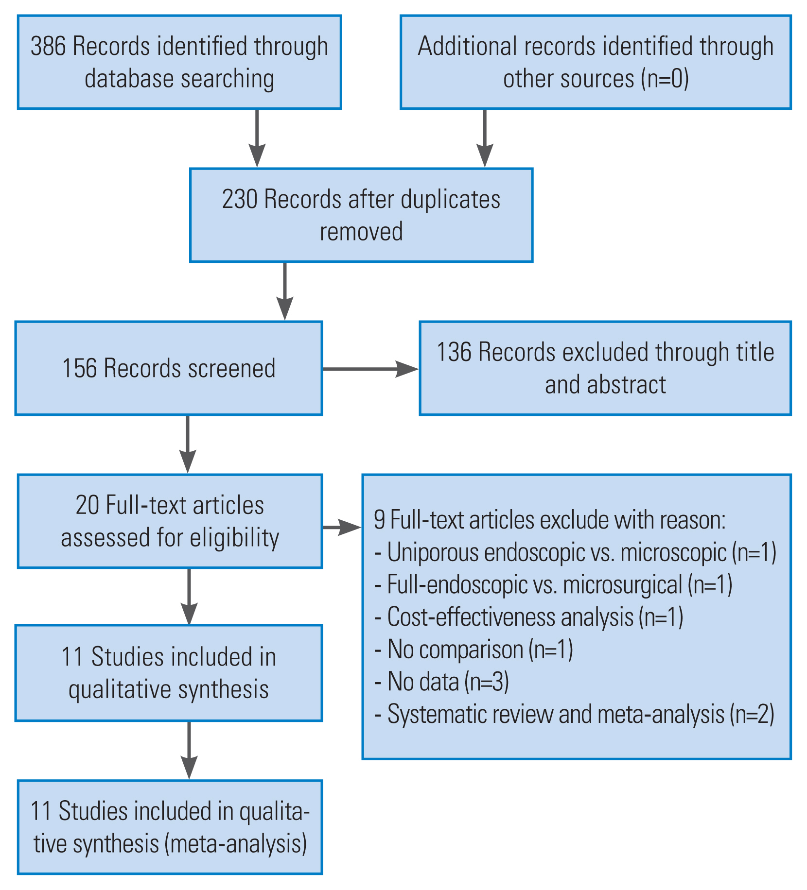
A total of 386 related documents were obtained through a preliminary search. Eleven articles were ultimately included [15,2130] (Fig. 1, Table 1) after rescreening by eliminating duplicate documents, reading titles and abstracts, and full text. There were 10 articles in English [15,2128,30] and one article in Chinese [29]. There were 930 patients, including 449 patients in UBE and 521 in MD.

2. Study characteristics and risk assessment

Only two of the included studies were randomized controlled trials [26,27] and the overall quality was moderate to high, according to the Cochrane system (Fig. 2). The remaining nine studies were nonrandomized controlled trials [15,2125,2830]. Risk assessment was conducted according to the RoBANS guidelines. Each study had a certain degree of bias. Table 2 shows the specific evaluation results.

3. Operation time

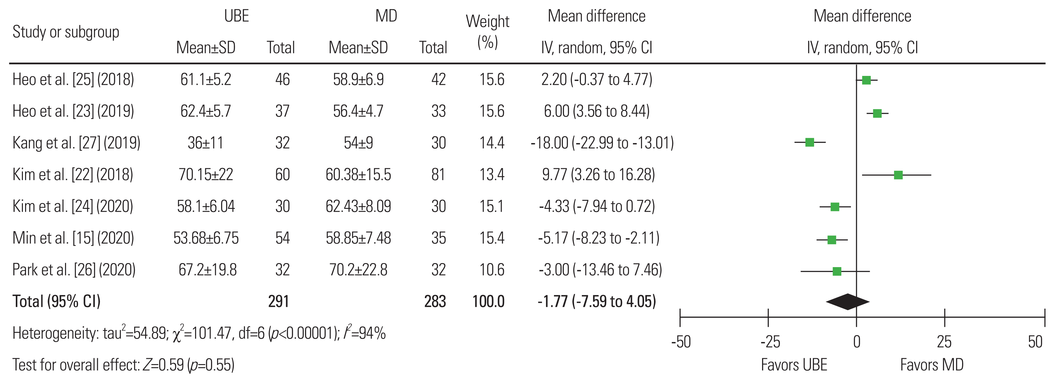
Seven studies were included to evaluate the operation time of the two surgery types with 291 patients in the UBE group and 283 in the MD group [15,2227]. There was statistical heterogeneity between the study groups (I2=94%). Thus, the random-effects model was used for statistical analysis and was grouped according to the different study types. There was no significant difference between the two (mean difference, −1.77; 95% CI, −7.59 to 4.05; p=0.55]. Therefore, in terms of operation time, a difference could not be identified between the two surgical methods (Fig. 3).

4. Blood loss

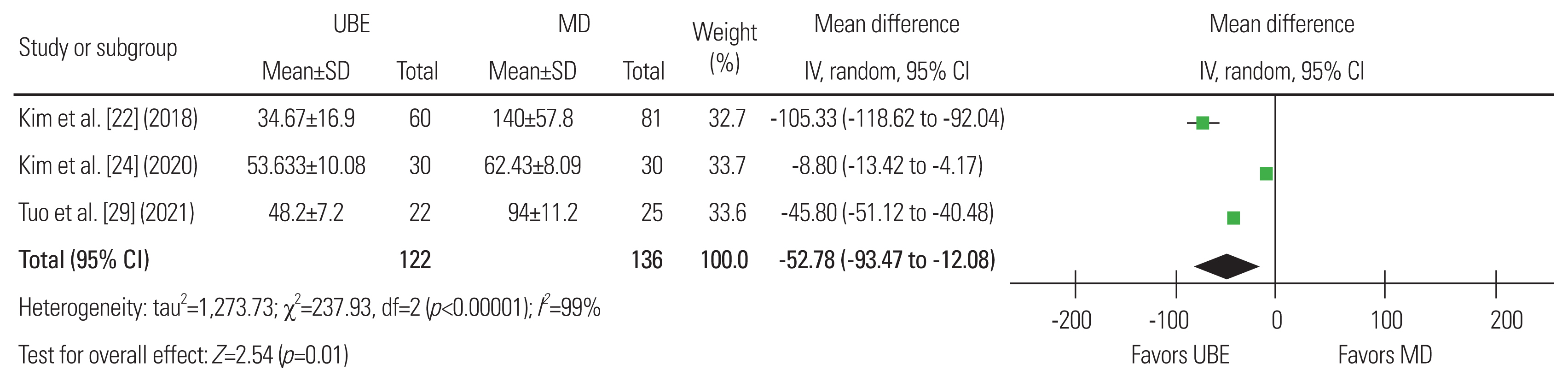
Three articles were included [22,24,29], with 112 patients in the UBE group and 136 in the MD group. The heterogeneity analysis showed significant heterogeneity (I2=99%) and a random-effects model was used. Results showed that blood loss was significantly different, favoring the UBE surgery group (p=0.01; mean difference, −52.78; 95% CI, −93.47 to −12.08). A mean difference of −52.78 mL (95% CI, −93.47 to −12.08 mL; p=0.01) concluding that UBE surgery results in less bleeding than MD surgery was revealed using these data for meta-analysis (Fig. 4).

5. Area of dural sac expansion

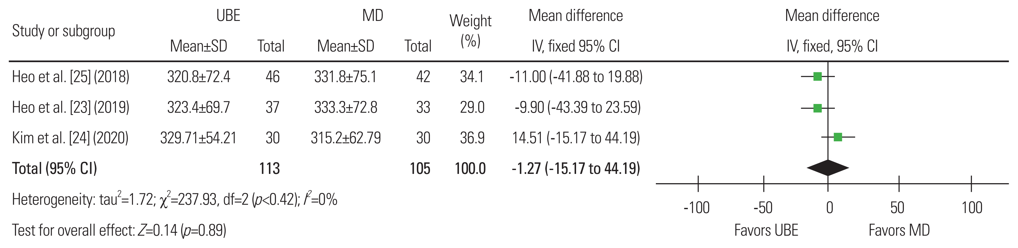
Three articles were included, with 113 patients in the UBE group and 105 in the MD group [2325]. The heterogeneity analysis showed that heterogeneity was not apparent (I2=0%) and the fixed-effects model was used. A mean difference of −1.27 (95% CI, −19.30, 16.77; p=0.89), suggesting that UBE surgery is similar in the degree of postoperative dural expansion to MD surgery, was shown using these data for meta-analysis (Fig. 5).

6. Postoperative complications

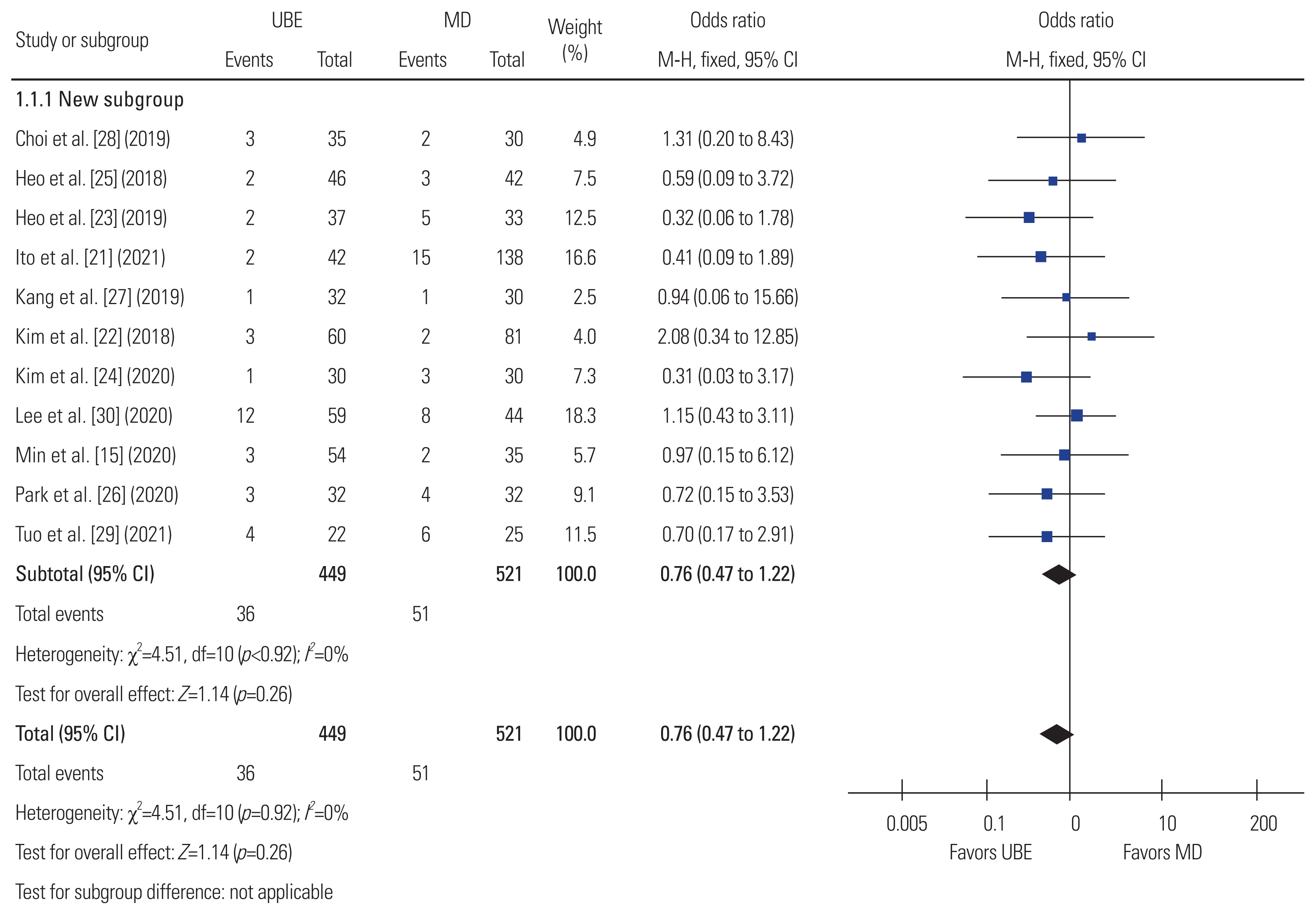
Eleven articles were included, with 449 patients in the UBE group and 521 in the MD group [15,2130]. The heterogeneity analysis showed that heterogeneity was not obvious (I2=0%). The fixed-effects model was used. A mean difference of 0.76 in postoperative complications (95% CI, 0.47 to 1.22; p=0.26), suggesting that UBE surgery is similar in terms of postoperative complications to MD surgery, was shown using these data for meta-analysis (Fig. 6).

7. Hospital stay

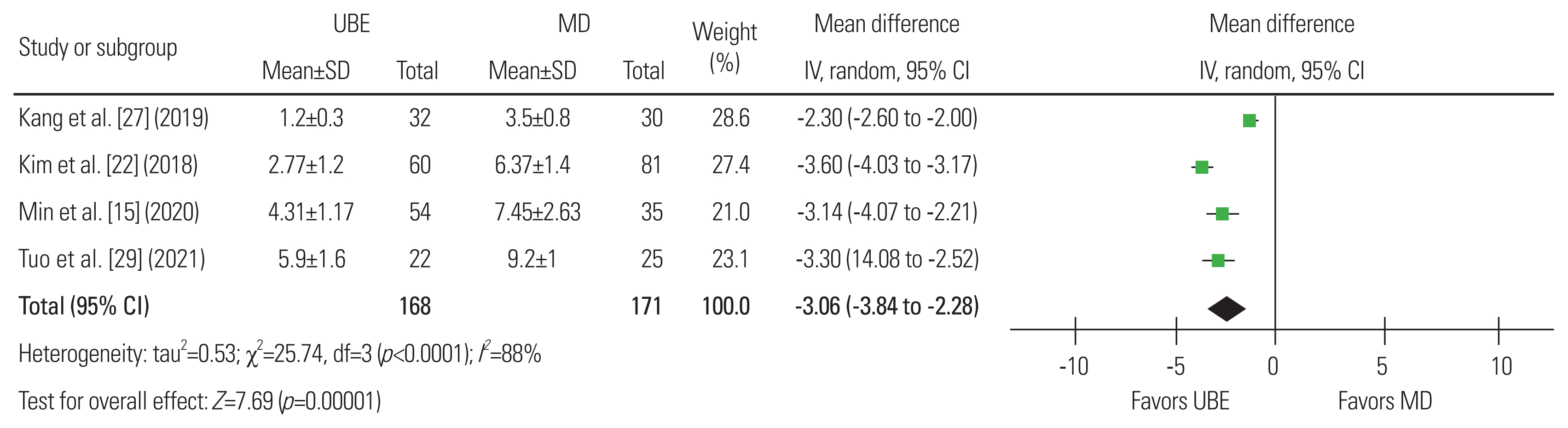
Four articles were included, with 168 patients in the UBE group and 171 in the MD group [15,22,27,29]. The analysis of heterogeneity showed significant heterogeneity (I2=88%), and a random-effects model was used. A meta-analysis of these data revealed a mean difference of −3.06 days (95% CI, −3.84 to 2.28 days; p<0.01), concluding that the UBE operation has a shorter hospital stay than the MD operation (Fig. 7).

8. C-reactive protein changes 1 week after surgery

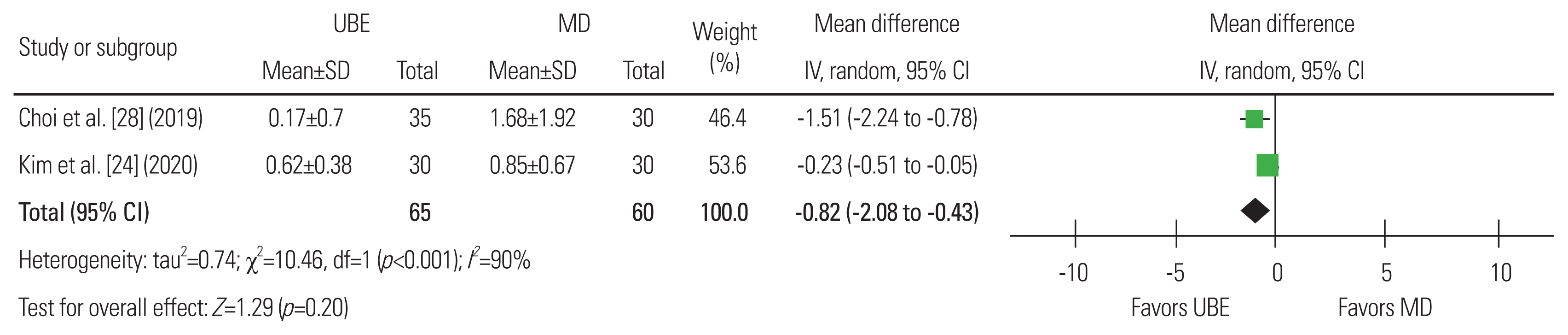
Two articles were included, with 65 patients in the UBE group and 60 in the MD group [24,28]. Significant heterogeneity was observed (I2=90%), and a random-effects model was used. A mean difference of −0.82 mg/L (95% CI, −2.08 to 0.43 mg/L; p=0.20), suggesting that UBE surgery is similar in terms of CRP value at 1 week after surgery to MD surgery, was shown using these data for meta-analysis (Fig. 8).

9. Visual Analog Scale score

Eight articles were included, with 597 patients in the UBE group and 586 in the MD group [15,2226,28,29]. Both the overall and group heterogeneity analyses showed mild heterogeneity (I2≤50%), and the fixed-effects model was used. A meta-analysis of these data revealed a mean difference of −0.04 in the VAS score (95% CI, −0.14 to 0.06 score; p=0.47). There was no significant difference between the two groups in terms of VAS for leg pain at the end of follow-up (more than 12 months) (Fig. 9).

10. Oswestry Disability Index score

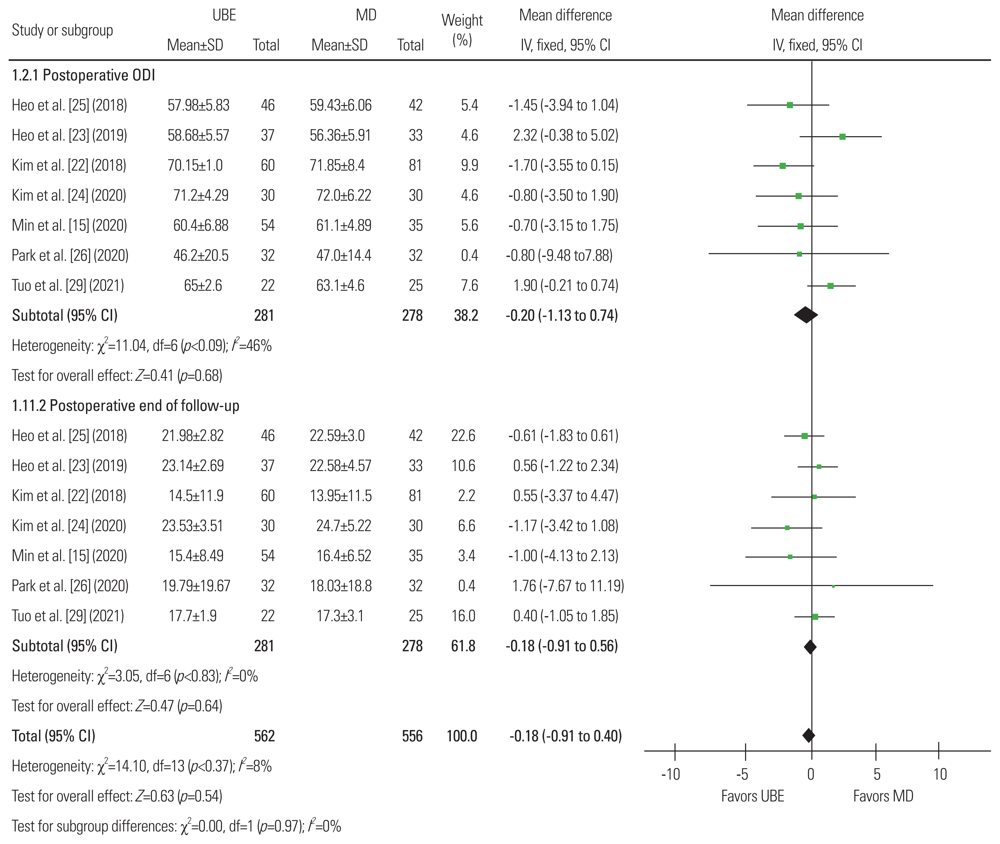
Seven articles were included, with 562 patients in the UBE group and 556 in the MD group [15,2226,29]. Both the overall and group heterogeneity analyses showed that heterogeneity was not obvious (I2<50%) and the fixed-effects model was used. A meta-analysis of these data revealed a mean difference of −0.18 in the ODI score (95% CI, −0.76 to 0.40; p=0.54). There was no difference between the ODI score at the preoperative ODI and the ODI score at the last follow-up (more than 12 months) (Fig. 10).

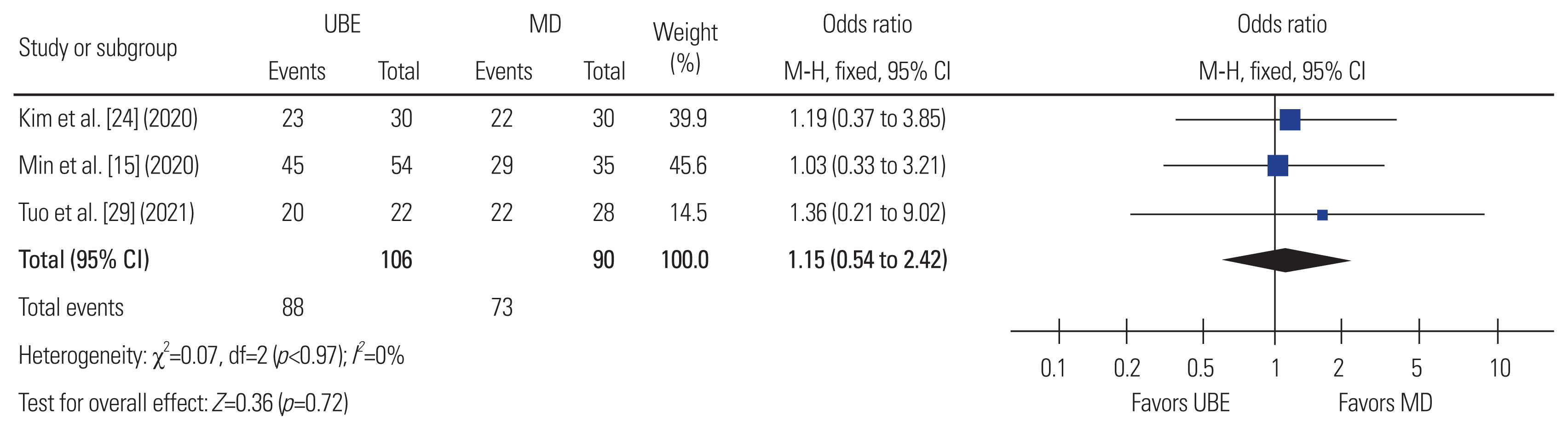
11. The patient satisfaction (the modified MacNab score)

Only three articles were included, with 106 patients in the UBE group and 90 in the MD group [15,24,29]. The heterogeneity analysis showed that the heterogeneity was not obvious (I2=0%) and the fixed-effects model was used. A meta-analysis of these data revealed a mean difference of −0.18 (95% CI, −0.76 to 0.40; p=0.54). Thus, it was not possible to determine a difference in patient satisfaction based on the postoperative modified MacNab score between the two types of surgery (Fig. 11).

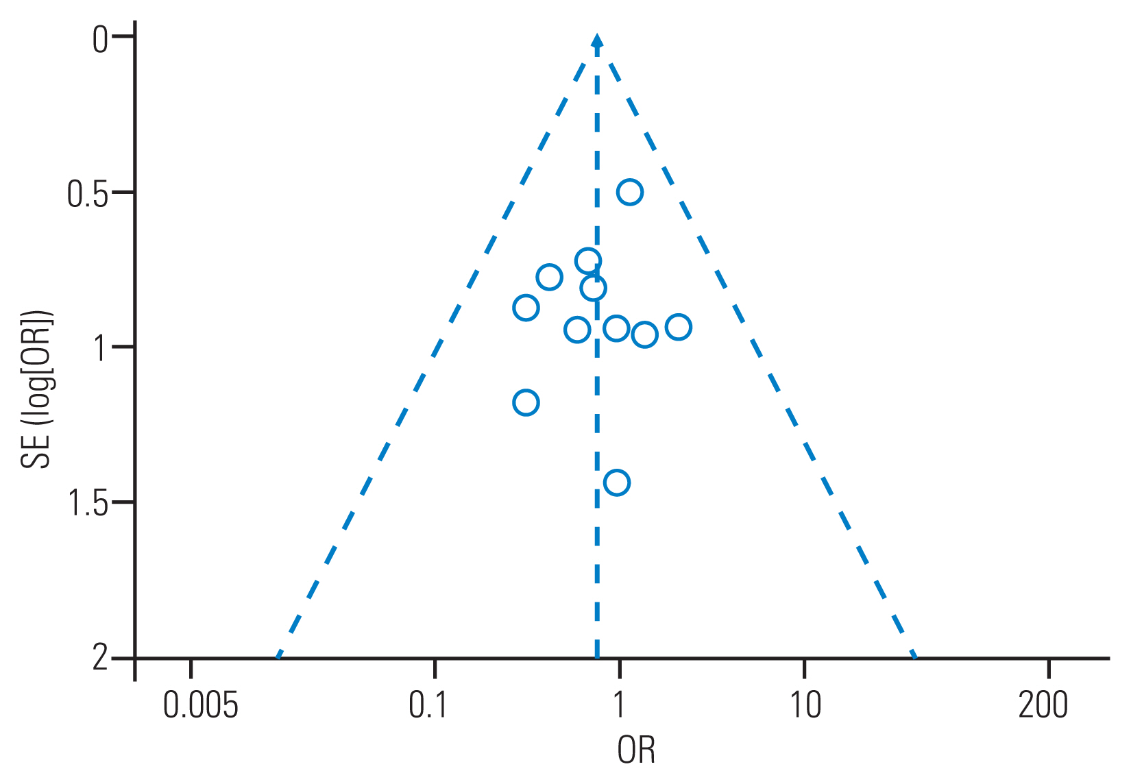
12. Publishing bias evaluation

The postoperative complications with the most included literature were selected for publication bias analysis, and a funnel chart was made. The 11 points in the figure were roughly symmetrically distributed, indicating that publication bias had a small effect on the results (Fig. 12).

Discussion

MD technology has always been the gold standard treatment for lumbar spinal stenosis [3,4]; however, it has certain limitations. It is limited in the scope of operation of the device and the field of view, which may lead to unsatisfactory decompression effects because this technique uses one incision. At the same time, the technology has a long learning curve, which makes it difficult for spinal surgeons to learn [510]. In recent years, the emerging UBE/BESS technology has used arthroscopic and conventional instruments. Through two channels, the instruments have the advantages of greater operating space and flexibility and a greater field of view. At the same time, it also has the advantages of less intraoperative bleeding and trauma; a smaller, shorter learning curve; increased safety and efficiency; and fewer complications. This technique has been applied to the treatment of lumbar spinal stenosis and has achieved satisfactory results [3133].
From this study, there was no significant difference between the two surgical procedures in terms of operation time, postoperative dural expansion area, complications, or the modified MacNab score. The two surgical techniques included complications, preoperative VAS for leg pain and last follow-up (>12 months) VAS for leg pain, and preoperative ODI score and last follow-up (>12 months) ODI score, and there were no significant differences in any of the aspects. However, a subgroup analysis was carried out due to heterogeneity, and results showed no significant differences. This indicates that these two techniques may be equally applicable to the treatment of lumbar spinal stenosis, are safe and effective, and have similar clinical effects.
The spinal canal decompression extent can be studied using magnetic resonance imaging to measure the dural mater area after surgery. Two minimally invasive surgical techniques were used to treat lumbar spinal stenosis after sufficient decompression, and spinal canal stenosis was significantly enlarged. UBE technique was more flexible and the decompression was more thorough although there was no significant difference between the two, especially for patients with severe lumbar central canal stenosis [34]. Pao et al. [11] showed that the average increase in the cross-sectional dural area after the UBE technique was 105.9±39.5 mm2, which corresponds to a preoperative increase of 201.9%±188.0%. Our comparison results also showed that the UBE technique has greatly improved after surgery. Research has shown that the technology has a short learning curve and can safely and effectively reduce pressure [11].
Serum creatine kinase (CK) and CRP have been used as parameters to assess muscle injury and acute inflammation, respectively. It was impossible to compare the two techniques in terms of surgical trauma due to the lack of postoperative CK data in this study. The results may have been affected by the large heterogeneity of the included studies although the results of this study showed that there were no significant differences in CRP 1 week after the operation. Choi and Kim [28] found that CK and CRP after the UBE technique were lower than those after the MD technique, indicating that the risk of muscle destruction and postoperative infection in UBE surgery was considerably smaller than that of the MD technique [24]. The UBE technique mainly causes less muscle damage through the intermuscular approach and the continuous saline can better flush the inflammatory debris.
The results of this study showed that the UBE technique has lower intraoperative blood loss and hospital stay than the MD technique. There were still significant differences between the two surgical techniques although the included studies were heterogeneous. The average length of hospital stay for patients treated with the UBE technique was 3.06 days shorter than that with the MD technique, likely due to UBE’s ability to minimize tissue damage and reduce operative muscle and ligament damage, intraoperative blood loss, and postoperative back pain recovery time [15,22]. This indicates that UBE technology may have some advantages over MD technology and patients undergoing UBE surgery may return to daily activities earlier. The stability of the spine is one of the factors that affects the patient’s early activities in the ground. Studies have shown that UBE technology causes minimal damage to the soft tissues and facet joints behind the spine. It can also safely and effectively decompress lumbar spinal stenosis and maintain segmental stability [11,35]. Pao et al. [11] showed that after adequate decompression of lumbar spinal stenosis treated with UBE technology, the contralateral facet joint preservation rate was 92.9% and that of the approach side was 84.2%. This may be another advantage of this technology.
This study has some limitations that need to be acknowledged. First, the best evidence for a systematic review is a randomized controlled trial. However, this systematic review only included two randomized controlled trials and the remaining nine were retrospective controlled studies, which may have distorted the results due to selection and recall bias. Second, the few included studies and the inconsistency of the results of the included studies indicate a high degree of variability in the results. Third, in the data included in this study for comparison, only the intraoperative blood loss and hospital stay were significantly different, and the heterogeneity showed obvious heterogeneity, which may be related to factors that depend on the operation situation and other variables. Further studies should be conducted with the same baseline and a larger sample size. The results of this study still have practical guiding significance despite the limitations.

Conclusions

There were no significant differences in the efficacy or safety of UBE and MD technology in the treatment of patients with lumbar spinal stenosis. However, UBE technology has less intraoperative bleeding and a shorter hospital stay. It can be well applied to lumbar spinal stenosis. It has a slight advantage and is a better surgical option than MD technology. More randomized controlled studies are needed to strengthen the current findings due to the inclusion of fewer studies.

Notes

Conflict of Interest

No potential conflict of interest relevant to this article was reported.

Funding

This study was supported by the National Natural Science Foundation of China (No. 81760231; No. 81160220); Natural Science Foundation of Ningxia Hui Autonomous Region (CN) (No. NZ17153; No. NZ11197); First-Class Discipline Construction Founded Project of NingXia Medical University and the School of Clinical Medicine (NXYLXK2017A05); and Natural Science Foundation of Ningxia Hui Autonomous Region (No. 2022AAC03398).

Author Contributions

Conception and design: Li Junjie, Yuan Haifeng; data acquisition: Li Junjie, Yin Jiheng, Liu Jun; analysis of data: Li Junjie, Yin Jiheng, Liu Jun; drafting of the manuscript: Li Junjie; critical revision: Li Junjie, Yin Jiheng, Liu Jun, Yuan Haifeng; obtaining funding: Lin haixiong, Yuan Haifeng; administrative support: Lin haixiong, Yuan Haifeng; and supervision: Yuan Haifeng.

Fig. 1
PRISMA (Preferred Reporting Items for Systematic reviews and Meta-Analyses) flow chart for the study search.
asj-2021-0527f1.jpg
Fig. 2
Judgement of the risk of bias included in the randomized controlled trial (RCT). (A) RCT risk of bias graph. (B) Risk of bias summary.
asj-2021-0527f2.jpg
Fig. 3
Forest plot of operation times for unilateral biportal endoscopy (UBE) vs. microscopic decompression (MD). SD, standard deviation; IV, inverse variance; CI, confidence interval; df, degrees of freedom.
asj-2021-0527f3.jpg
Fig. 4
Forest plot of estimated blood loss for unilateral biportal endoscopy (UBE) vs. microscopic decompression (MD). SD, standard deviation; IV, inverse variance; CI, confidence interval; df, degrees of freedom.
asj-2021-0527f4.jpg
Fig. 5
Forest plot of the area of dural sac expansion for unilateral biportal endoscopy (UBE) vs. microscopic decompression (MD). SD, standard deviation; IV, inverse variance; CI, confidence interval; df, degrees of freedom.
asj-2021-0527f5.jpg
Fig. 6
Forest plot of complications associated with unilateral biportal endoscopy (UBE) vs. microscopic decompression (MD). M-H, Mantel–Haenszel; CI, confidence interval; df, degrees of freedom.
asj-2021-0527f6.jpg
Fig. 7
Forest plot of lengths of hospital stay for unilateral biportal endoscopy (UBE) vs. microscopic decompression (MD). SD, standard deviation; IV, inverse variance; CI, confidence interval; df, degrees of freedom.
asj-2021-0527f7.jpg
Fig. 8
Forest plot of CRP for the unilateral biportal endoscopy (UBE) vs. microscopic decompression (MD). SD, standard deviation; IV, inverse variance; CI, confidence interval; df, degrees of freedom.
asj-2021-0527f8.jpg
Fig. 9
Subgroup analysis forest plot of preoperative and postoperative end of follow-up (more than 12 months) Visual Analog Scale (VAS) for leg pain for the unilateral biportal endoscopy (UBE) vs. microscopic decompression (MD). SD, standard deviation; IV, inverse variance; CI, confidence interval; df, degrees of freedom.
asj-2021-0527f9.jpg
Fig. 10
Subgroup analysis forest plot of preoperative and postoperative end of follow-up (more than 12 months) Oswestry Disability Index (ODI) scores for unilateral biportal endoscopy (UBE) vs. microscopic decompression (MD). SD, standard deviation; IV, inverse variance; CI, confidence interval; df, degrees of freedom.
asj-2021-0527f10.jpg
Fig. 11
Forest plot of patient satisfaction (the modified MacNab score) for unilateral biportal endoscopy (UBE) vs. microscopic decompression (MD). M-H, Mantel–Haenszel; CI, confidence interval; df, degrees of freedom.
asj-2021-0527f11.jpg
Fig. 12
Funnel diagram of complications in the two surgical methods. OR, odds ratio; SE, standard error.
asj-2021-0527f12.jpg
Table 1
Characteristics of included studies
Study Design Country No. of patients (UBE/MD) Age (yr) (UBE/MD) Sex (M:F), (UBE/MD) No. of levels Complications Outcomes Final follow-up (mo)
Min et al. [15] (2020) Case control study Korea 54/35 65.74±10.52/66.74±7.96 27:27/19:16 All single level UBE: dural tear (n=2), postoperative epidural hematoma (n=1); MD: dural tear (n=1), postoperative epidural hematoma (n=1) VAS for back and leg; ODI; operative time; modified Macnab criteria; complication UBE: 27.2±5.4
MD: 31.5±7.3
Ito et al. [21] (2021) Single-center, retrospective analysis Japan 42/139 66.3±12.3/65.0±11.1 28:14/71:68 All single level UBE: dural injury (n=2); MD: hematoma (n=5), dural injury (n=8), reoperation (n=2) VAS for back and leg; ODI; operative time; bone resection area; facet; preservation rates; EQ-5D questionnaire; complication UBE: 6.7±0.6
MD: 6.9±0.8
Kim et al. [22] (2018) Multicenter, retrospective analysis Korea 60/81 46.60±14.18/54.22±20.21 37:23/24:57 All single level UBE: change the surgical plan (n=3); MD: cerebrospinal fluid leakage (n=1), cerebrospinal fluid leakage and infection (n=1) VAS for back and leg; ODI; operative time; complication; estimated blood loss; hospital stay; MacNab score; complications UBE: 12.60±1.03
MD: 12.84±1.30
Heo et al. [23] (2019) Retrospective analysis of prospectively Korea 37/33 66.7±9.4/63.4±11.1 15:22/12:21 All single level UBE: durotomy (n=1), postoperative hematoma (n=1) MD: durotomy (n=2), transient weakness (n=1), postoperative hematoma (n=2) VAS for back and leg; ODI; operative time; dura expansion; angle of facetecomy; complication UBE: 12.5±3.3
MD: 12.5±3.3
Kim et al. [24] (2020) Retrospective study Korea 30/30 64.23±5.26/66.20±6.01 13:17/12:18 All single level UBE: cerebrospinal fluid leak (n=1); MD: cerebrospinal fluid leak (n=2), surgical site infection (n=1) VAS; ODI; operative time; dura expansion; estimated blood loss; serum creatine kinase; CRP; modified MacNab score; complication UBE: 12
MD: 12
Heo et al. [25] (2018) A case control prospective study Korea 46/42 65.8±8.9/63.6±10.5 18:28/16:26 All single level UBE: durotomy (n=1), postoperative hematoma (n=1); MD: durotomy (n=1), postoperative hematoma (n=2) VAS for back and leg; ODI; operative time; cross-sectional area of the dura; complication UBE: 14.5±2.3
MD: 14.5±2.3
Park et al. [26] (2020) Randomized controlled trial Korea 32/32 66.2 (41–80)/ 67.1 (45–79) 13 :19/18 :14 All single level UBE: incidental durotomy (n=2), postoperative epidural hematoma (n=1); MD: incidental durotomy (n=2), postoperative epidural hematoma (n=1), revision surgery due to recurrent pain (n=1) VAS for back and leg; ODI; operative time; complication; EQ-5D score; hospital stay; postoperative drainage; serum creatine phosphokinase UBE: 12
MD: 12
Kang et al. [27] (2019) A prospective randomized comparative study Korea 32/30 65.1±8.6/67.2±9.5 18:14/14:16 All single level UBE: revision (n=1); MD: revision (n=1) Operative time; hospital stay; Hemovac drain output; complication UBE: 6
MD: 6
Choi et al. [28] (2019) Retrospective study Korea 35/30 65.4±11.8/65.2±12.0 14:21/17:13 UBE: single level (24), multiple levels (11); MD: single level (15), multiple level (15) UBE: dural tear (n=2), root injury (n=1); MD: dural tear (n=2) VAS for back and leg; ODI; CRP; complication; postoperative hemoglobin; transfusion UBE: 6
MD: 6
Tuo et al. [29] (2021) Retrospective analyzed China 22/25 59.1±11.7/58.3±8.7 12:10/11:14 All single level UBE: postoperative epidural hematoma (n=1), dura tear (n=3) MD: postoperative epidural hematoma (n=1), dura tear (n=5) VAS; ODI; operative time; hospital stay; intraoperative blood loss; complication; modified MacNab criteria UBE: 12
MD: 12
Lee et al. [30] (2020) Retrospective Korea 59/44 69.64±11.05/69.11±8.58 30:29/16:28 All single level UBE: dura tear (n=6), infection (n=2), postoperative epidural hematoma (n=1), incomplete decompression (n=2), others (n=1); MD: dura tear (n=4), infection (n=2), postoperative epidural hematoma (n=1), others (n=1) VAS for back and leg; ODI; operative time; drainage volume; hospital stay; complication UBE: 3
MD: 3

Values are presented as number, mean±standard deviation, or mean (range), otherwise stated.

UBE, unilateral biportal endoscopy; MD, microscopic decompression; M, male; F, female; VAS, Visual Analog Scale; ODI, Oswestry Disability Index; EQ-5D, European Quality of Life-5 Dimensions; CRP, C-reactive protein.

Table 2
Quality evaluation according to the Risk of Bias Assessment of Non-randomized Studies Scale
Study Selection of participants Confounding variables Measurement of exposure Blinding of outcome assessment Incomplete outcome data Selective reporting outcome
Min et al. [15] (2020) Low risk Unclear risk Low risk High risk Low risk Low risk
Ito et al. [21] (2021) Low risk Low risk Low risk High risk Low risk Low risk
Kim et al. [22] (2018) Low risk Low risk Low risk High risk Low risk Low risk
Heo et al. [23] (2019) Low risk High risk Unclear risk High risk Low risk Low risk
Lee et al. [30] (2020) Low risk Unclear risk Low risk High risk Low risk Low risk
Choi et al. [28] (2019) Low risk High risk Unclear risk High risk Low risk Low risk
Tuo et al. [29] (2021) Low risk Low risk Low risk High risk Low risk Low risk
Kim et al. [24] (2020) Low risk High risk Low risk High risk Low risk Low risk
Heo et al. [25] (2018) Low risk Low risk Low risk High risk Low risk Low risk

References

1. Kim M, Kim HS, Oh SW, et al. Evolution of spinal endoscopic surgery. Neurospine 2019 16:6–14.
crossref pmid pmc pdf
2. Pranata R, Lim MA, Vania R, July J. Biportal endoscopic spinal surgery versus microscopic decompression for lumbar spinal stenosis: a systematic review and meta-analysis. World Neurosurg 2020 138:e450–8.
crossref pmid
3. Eum JH, Heo DH, Son SK, Park CK. Percutaneous biportal endoscopic decompression for lumbar spinal stenosis: a technical note and preliminary clinical results. J Neurosurg Spine 2016 24:602–7.
crossref pmid
4. Takahashi H, Aoki Y, Saito J, et al. Unilateral laminectomy for bilateral decompression improves low back pain while standing equally on both sides in patients with lumbar canal stenosis: analysis using a detailed visual analogue scale. BMC Musculoskelet Disord 2019 20:100.
crossref pmid pmc pdf
5. Uehara M, Takahashi J, Hashidate H, et al. Comparison of spinous process-splitting laminectomy versus conventional laminectomy for lumbar spinal stenosis. Asian Spine J 2014 8:768–76.
crossref pmid pmc
6. Mobbs RJ, Li J, Sivabalan P, Raley D, Rao PJ. Outcomes after decompressive laminectomy for lumbar spinal stenosis: comparison between minimally invasive unilateral laminectomy for bilateral decompression and open laminectomy: clinical article. J Neurosurg Spine 2014 21:179–86.
pmid
7. Ko S, Oh T. Comparison of bilateral decompression via unilateral laminotomy and conventional laminectomy for single-level degenerative lumbar spinal stenosis regarding low back pain, functional outcome, and quality of life: a randomized controlled, prospective trial. J Orthop Surg Res 2019 14:252.
crossref pmid pmc pdf
8. Mobbs R, Phan K. Minimally invasive unilateral laminectomy for bilateral decompression. JBJS Essent Surg Tech 2017 7:e9.
crossref pmid pmc
9. Phan K, Mobbs RJ. Minimally invasive versus open laminectomy for lumbar stenosis: a systematic review and meta-analysis. Spine (Phila Pa 1976) 2016 41:E91–100.
pmid
10. Ulrich NH, Burgstaller JM, Gravestock I, et al. Outcome of unilateral versus standard open midline approach for bilateral decompression in lumbar spinal stenosis: is “over the top” really better?: a Swiss prospective multicenter cohort study. J Neurosurg Spine 2019 31:236–45.
crossref
11. Pao JL, Lin SM, Chen WC, Chang CH. Unilateral biportal endoscopic decompression for degenerative lumbar canal stenosis. J Spine Surg 2020 6:438–46.
crossref pmid pmc
12. Kim JE, Choi DJ, Park EJJ, et al. Biportal endoscopic spinal surgery for lumbar spinal stenosis. Asian Spine J 2019 13:334–42.
crossref pmid pmc pdf
13. Wang MC, Yu KY, Zhang JG, Wang YP. Progression and clinical application in unilateral biportal endoscopic. Zhonghua Wai Ke Za Zhi 2020 58:892–6.
pmid
14. Pairuchvej S, Muljadi JA, Ho JC, Arirachakaran A, Kongtharvonskul J. Full-endoscopic (bi-portal or uni-portal) versus microscopic lumbar decompression laminectomy in patients with spinal stenosis: systematic review and meta-analysis. Eur J Orthop Surg Traumatol 2020 30:595–611.
crossref pmid pdf
15. Min WK, Kim JE, Choi DJ, Park EJ, Heo J. Clinical and radiological outcomes between biportal endoscopic decompression and microscopic decompression in lumbar spinal stenosis. J Orthop Sci 2020 25:371–8.
crossref pmid
16. Eun SS, Eum JH, Lee SH, Sabal LA. Biportal endoscopic lumbar decompression for lumbar disk herniation and spinal canal stenosis: a technical note. J Neurol Surg A Cent Eur Neurosurg 2017 78:390–6.
crossref pmid
17. Kim W, Kim SK, Kang SS, Park HJ, Han S, Lee SC. Pooled analysis of unsuccessful percutaneous biportal endoscopic surgery outcomes from a multi-institutional retrospective cohort of 797 cases. Acta Neurochir (Wien) 2020 162:279–87.
crossref pmid pdf
18. Chen T, Zhou G, Chen Z, Yao X, Liu D. Biportal endoscopic decompression vs. microscopic decompression for lumbar canal stenosis: a systematic review and meta-analysis. Exp Ther Med 2020 20:2743–51.
crossref pmid pmc
19. Higgins JP, Thomas J, Chandler J, et al. Cochrane handbook for systematic reviews of interventions. 2nd ed. Chichester: John Wiley & Sons Ltd; 2019.

20. Kim SY, Park JE, Lee YJ, et al. Testing a tool for assessing the risk of bias for nonrandomized studies showed moderate reliability and promising validity. J Clin Epidemiol 2013 66:408–14.
crossref pmid
21. Ito Z, Shibayama M, Nakamura S, et al. Clinical Comparison of unilateral biportal endoscopic laminectomy versus microendoscopic laminectomy for single-level laminectomy: a single-center, retrospective analysis. World Neurosurg 2021 148:e581–8.
crossref pmid
22. Kim SK, Kang SS, Hong YH, Park SW, Lee SC. Clinical comparison of unilateral biportal endoscopic technique versus open microdiscectomy for single-level lumbar discectomy: a multicenter, retrospective analysis. J Orthop Surg Res 2018 13:22.
crossref pmid pmc pdf
23. Heo DH, Lee DC, Park CK. Comparative analysis of three types of minimally invasive decompressive surgery for lumbar central stenosis: biportal endoscopy, uniportal endoscopy, and microsurgery. Neurosurg Focus 2019 46:E9.
crossref
24. Kim HS, Choi SH, Shim DM, Lee IS, Oh YK, Woo YH. Advantages of new endoscopic unilateral laminectomy for bilateral decompression (ULBD) over conventional microscopic ULBD. Clin Orthop Surg 2020 12:330–6.
crossref pmid pmc pdf
25. Heo DH, Quillo-Olvera J, Park CK. Can percutaneous biportal endoscopic surgery achieve enough canal decompression for degenerative lumbar stenosis?: prospective case-control study. World Neurosurg 2018 120:e684–9.
crossref pmid
26. Park SM, Park J, Jang HS, et al. Biportal endoscopic versus microscopic lumbar decompressive laminectomy in patients with spinal stenosis: a randomized controlled trial. Spine J 2020 20:156–65.
crossref pmid
27. Kang T, Park SY, Kang CH, Lee SH, Park JH, Suh SW. Is biportal technique/endoscopic spinal surgery satisfactory for lumbar spinal stenosis patients?: a prospective randomized comparative study. Medicine (Baltimore) 2019 98:e15451.
pmid pmc
28. Choi DJ, Kim JE. Efficacy of biportal endoscopic spine surgery for lumbar spinal stenosis. Clin Orthop Surg 2019 11:82–8.
crossref pmid pmc pdf
29. Wei T, Lin Z, Desen L, Liuwei G, Xiaokai X, Hong C. Preliminary study of unilateral biportal endoscopic treatment of lumbar spinal stenosis. Chin J Min Inv Surg 2021 21:56–60.

30. Lee JH, Cho SI, Chough CK. Surgical outcomes of microscopic versus unilateral biportal endoscopic decompression for lumbar spinal stenosis: a preliminary report. J Korean Soc Geriatr Neurosurg 2020 16:42–6.
crossref pdf
31. Park SM, Kim HJ, Kim GU, et al. Learning curve for lumbar decompressive laminectomy in biportal endoscopic spinal surgery using the cumulative summation test for learning curve. World Neurosurg 2019 122:e1007–13.
crossref pmid
32. Lin GX, Huang P, Kotheeranurak V, et al. A systematic review of unilateral biportal endoscopic spinal surgery: preliminary clinical results and complications. World Neurosurg 2019 125:425–32.
crossref pmid
33. Ahn JS, Lee HJ, Choi DJ, Lee KY, Hwang SJ. Extraforaminal approach of biportal endoscopic spinal surgery: a new endoscopic technique for transforaminal decompression and discectomy. J Neurosurg Spine 2018 28:492–8.
crossref pmid
34. Kim N, Jung SB. Percutaneous unilateral biportal endoscopic spine surgery using a 30-degree arthroscope in patients with severe lumbar spinal stenosis: a technical note. Clin Spine Surg 2019 32:324–9.
pmid pmc
35. Kim JE, Choi DJ. Clinical and radiological outcomes of unilateral biportal endoscopic decompression by 30° arthroscopy in lumbar spinal stenosis: minimum 2-year follow-up. Clin Orthop Surg 2018 10:328–36.
crossref pmid pmc pdf


ABOUT
ARTICLE CATEGORY

Browse all articles >

BROWSE ARTICLES
EDITORIAL POLICY
FOR CONTRIBUTORS
Editorial Office
Department of Orthopedic Surgery, Asan Medical Center, University of Ulsan College of Medicine
88, Olympic-ro 43-gil, Songpa-gu, Seoul 05505, Korea
Tel: +82-2-3010-3530    Fax: +82-2-3010-8555    E-mail: asianspinejournal@gmail.com                
Korean Society of Spine Surgery
27, Dongguk-ro, Ilsandong-gu, Goyang-si 10326, Korea
Tel: +82-31-966-3413    Fax: +82-2-831-3414    E-mail: office@spine.or.kr                

Copyright © 2024 by Korean Society of Spine Surgery.

Developed in M2PI

Close layer
prev next