Introduction
Ponticulus posticus has recently been reported to predispose a catastrophic complication such as a vertebral artery injury, during the placement of lateral mass screws into the atlas. The Latin meaning of ponticulus posticus is "little posterior bridge"1, which describes a malformed bony bridge between the posterior portion of the superior articular process and the posterolateral portion of the superior margin of the posterior arch of the atlas. Ponticulus posticus forms the arcuate foramen that contains the vertebral artery and the suboccipital nerve and has been known to be a possible cause of posterior circulation strokes2 and migraines3. This condition had not been a matter of concern for spine surgeons until its surgical significance in the placement of screws into the lateral mass of the Atlas was recently reported1. Young et al.4 reported that mistaking the ponticulus posticus for just a broad posterior arch of the atlas during C1 lateral mass screw placement could cause injury to the vertebral artery. Considering the catastrophic complication this could possibly cause, we need to understand the three-dimensional morphologic features and the prevalence of this anomaly.
Prevalence of Ponticulus Posticus has been reported to be between 5.1% and 37.8% in the Western population3-5. We, however, could find only one report1 on its prevalence or morphologic characteristics in an Asian population. Therefore, we investigated the prevalence and morphologic features of ponticulus posticus in Koreans.
Materials and Methods
This study was approved by the institutional review board of our institution. First, we retrieved 1-mm interval cervical CT scan images of 225 consecutive patients over the age of eighteen taken in a large metropolitan teaching hospital. In most of those patients, CT scanning was ordered by orthopaedic surgeons or neurosurgeons for evaluation of cervical spine problems. There were 146 men and 79 women, and the average age was 45 (range, 18 to 87). All the images were retrieved in digital format from a picture archiving and communication system (PACS) server. Using personal computer-based software (V-works; Cybermed, Korea), these images were reconstructed into three-dimensional images, and carefully inspected for the presence and types of ponticuli.
Because the 225 patients examined above had cervical spine problems, we could not be sure whether the prevalence of ponticuli in these patients was an accurate representation of this condition in the normal Korean population. We therefore examined digital lateral cephalometric head radiographs of 315 consecutive patients over the age of eighteen, which were taken in the department of dentistry of the same hospital for evaluation of dental conditions, facial patterns, and jaw relationships, regardless of presence or absence of any cervical symptoms or headaches. Three patients with poor visualization of the posterior arch of the atlas due to overlapping of the mastoid process or the occiput were excluded. Of the 312 patients examined, 117 were men and 195 were women. The average age was 28 (range, 18 to 69). In each radiograph the presence of a ponticulus posticus and whether it was complete or partial were carefully inspected.
Statistical analysis was conducted with the chi-square test, using SPSS statistical package (version 12.0; SPSS Inc., Chicago, IL, USA). The level of significance was set at p<0.05.
Results
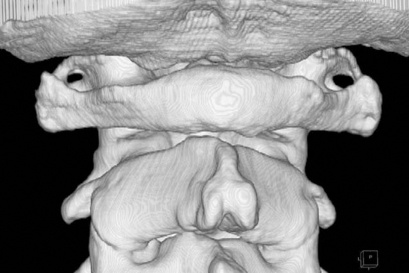
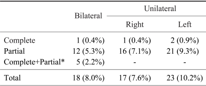
Analysis of the 225 three-dimensional CT scans revealed ponticulus posticus in 58 patients, comprising 26% (Table 1). The ponticulus was bilateral in 18 patients (8%); on the right side alone in 17 patients (8%); and on the left side alone in 23 patients (10%). The difference in frequency between the right and left sides was not statistically significant (p=0.45). Among the 76 ponticuli posticus observed on one or both sides in 58 patients, nine were complete and sixty-seven were incomplete (Fig. 1). There was no significant difference in the prevalence between men (37 out of 146, 25%) and women (21 out of 79, 27%; p=0.84). We also observed ponticulus lateralis (Fig. 2), which is a bony bridge between the lateral aspect of the superior articular process and the transverse process, in 14 patients (6%). All of them except one were unilateral, on the right side alone in 4 patients (2%) and on the left side alone in 9 patients (4%).
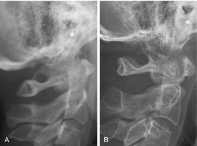
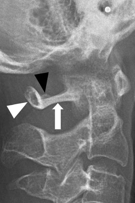
Analysis of the digital lateral cephalometric head radiographs of 312 patients revealed 14 complete and 30 incomplete ponticuli posticus (Fig. 3). Thus, the overall prevalence of ponticulus posticus in this patient population was 14%, comprising 4% complete and 10% incomplete. This was significantly smaller than the prevalence in the cervical spine patients in whom CT scans were analyzed (p=0.0007). There was no significant difference in the prevalence between men (19 out of 117, 16%) and women (25 out of 195, 13%; p=0.40). In another 20 patients (6%), spicules resembling partial ponticulus were found. Their sizes were not so large that we could not classify them as ponticuli posticus (Fig. 4). However, they might be a mild form of partial ponticulus posticus.
Discussion
Ponticulus posticus has become an important anomaly of the atlas, as the use of lateral mass screws for the fixation of the atlas has become common. Because putting this screw in the classical entry point at the junction of the posterior arch and the lateral mass6,7 causes significant bleeding from the epidural plexus and can possibly cause irritation to the C2 nerve root resulting in occipital neuralgia, some surgeons recommend placing the screw higher, starting in the posterior aspect of the posterior arch8. A broad posterior arch of the atlas is the best indication for this modified screw trajectory. However, in patients having a ponticulus posticus, it can be mistaken for a broad posterior arch, and the surgeon may put the screw into the ponticulus posticus. This could result in an injury to the vertebral artery which is trapped in the arcuate foramen and can scarcely escape injury by the probe, drill bit, tap, and/or screw. Vertebral artery injury may not cause any symptoms if the patient and the surgeon are lucky, but otherwise, it can lead to stroke or even death by thrombosis, embolism, or arterial dissection. Considering the significance of this grave complication, we need to investigate the morphological characteristics and prevalence of the ponticulus posticus.
In the western population, the prevalence of ponticulus posticus has been reported to be between 5.1% and 37.8%3-5. However, as far as we know, the only study on its prevalence in an Asian population is from India1. This study analyzed north Indians, many of whom are presumed to be Aryan. Therefore, we examined the three-dimensional CT scan images of Korean people. We were astonished that as much as 26% of the patient population had ponticulus posticus, because we had not noticed through our daily practice that its prevalence was so high. Most of these patients took a CT scan because of cervical spine problems, and patients who had upper cervical problems including congenital anomalies were not uncommon among them. Therefore, we presumed that these patients did not represent the whole population. So we then examined lateral cephalometric head radiographs of dental patients which were taken regardless of presence or absence of cervical symptoms. Surprisingly, this examination revealed 14% prevalence of ponticulus posticus. We think that the statistically significant difference (p=0.0007) in prevalence between the two groups was not only due to different patient populations but also influenced by the different diagnostic values of CT scans and radiographs. In any case, we found that ponticulus posticus is not a rare anomaly in Koreans, just as it is not in the Western population. Therefore, lateral radiographs should be carefully examined to check for the presence of this anomaly before screw placement in the lateral mass of the atlas in order to avoid vertebral artery injury.
In addition, we found that 20 patients (7%) had small spicules resembling mild partial ponticuli on radiographs (Fig. 4). We did not classify them as partial ponticuli because the spicules were not large, but they might have been a mild form of partial ponticulus posticus. Importantly, in these patients, the lateral portion of the posterior arch tended to be thin and low while the thickness of the posterior portion tended to look normal (Fig. 4). If a surgeon did not take a careful look at this morphological characteristic seen on the radiograph and put a screw in the posterior arch, it could result in a vertebral artery injury or fracture or weakness of this portion.
Examination of the three-dimensional CT images revealed a various spectra of shapes and sizes of the ponticulus posticus. We also found that even the ponticulus lateralis was not uncommon. This wide variation of shapes, sizes and location of the ponticuli seems to be natural, considering they are normal occurrence in the quadrupeds and act as an additional extension for the attachment of the posterior atlantooccipital membrane2. In any case, ponticulus posticus can have a wide variation in shapes and sizes and we cannot ascertain its three dimensional morphology on each side of an individual patient without three-dimensional CT images. Therefore, when the ponticulus posticus is observed or suspected on the lateral radiograph of a patient who requires lateral mass screw placement in the atlas, we may have two options. One is to put the screws under the posterior arch in the classic way6,7. If placing screws in the posterior arch8 is desired, we recommend taking a three-dimensional CT scan and checking the size and shape of the ponticulus on each side in order to avoid vertebral artery injury. Since these patients will require screw fixation at other levels of the cervical spine or the occiput, CT scans will be helpful planning for placement of these screws as well.
Conclusions
The ponticulus posticus is not an uncommon anomaly in Korean people, though its prevalence in other Asian races remains unknown. Therefore, the presence of this anomaly should be carefully ruled out on lateral radiographs before screw placement in the lateral mass of the atlas in order to avoid vertebral artery injury. Because ponticulus posticus has a wide spectra of shapes and sizes which cannot be exactly determined using other studies, we recommend taking a three-dimensional CT scan when a ponticulus posticus is suspected or observed on the radiographs of a patient in whom lateral mass screw placement in the posterior arch of the atlas is planned.