Introduction
The long-term effects of instrumented spinal fusion on adjacent motion segments have become an issue of increasing concern. Many clinical and biomechanical studies have examined the degeneration or disease adjacent to the instrumented fusion segments [1-4]. A decrease in the number of mobile segments can cause increased stress in the unfused segments, predisposing them to early degeneration. Some authors have suggested that the thoracolumbar segments should be included to reduce the level of stress when the upper lumbar segments are fused. Furthermore, proximal junctional problems can occur more frequently for fusions ending at T11 to L2 compared to those endings at T10 or above [2]. In addition, extending the instrumentation to T10 or proximally might offer relative protection to the adjacent segments due to the increased stability afforded by the rib cage [2]. Freccero and Donnovan [5] described a rare case of adjacent segment degeneration at T1-T2 after cervicothoracic fusion (C5-T1). They suggested that fusion across the cervicothoracic junction might prevent further degeneration at levels below the constructs because the thoracic spine is stabilized by the ribs [5]. However, our patient developed T9-T10 thoracic disc herniation after T10-S1 fusion despite the T9-T10 stabilization by the rib cage.
To our knowledge, there are no reports of thoracic disc herniation with myelopathy of the adjacent segments at the T9-10. We describe a case of thoracic disc herniation of the adjacent segments fused to T10-S1 with rapidly progressing myelopathy.
Case Report
A 66-year-old woman suddenly developed motor impairment of both lower extremities and sensory impairment of the left lower extremity with one day duration. Three years earlier, she had undergone instrumented fusion from T10-S1 at another institution secondary to degenerative lumbar kyphosis and spinal stenosis. On this occasion, she initially presented at another hospital, where she denied any obvious trauma, and was admitted for observation after the radiographs. The motor impairment of both lower extremities deteriorated gradually over 2 days, which prevented her from standing without assistance. She presented at our institution 6 days after the onset of symptoms.
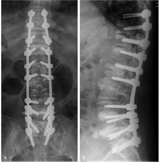
The physical examination revealed mild tenderness over the thoracolumbar area. The neurological examination showed positive bilateral Babinski reflexes, motor impairment both lower extremities (grade 1) on the manual muscle test scale, last normal sensory level at T10 with hyposthesia at T11 onwards with preservation of the anal wink and tone, and no bowel/bladder incontinence. The preoperative ASIA score for cervical myelopathy was calculated with a score of 60 on motor. The plain lumbar radiographs revealed instrumental fusion from S1 to T10 and narrowing of the intervertebral disc at T9-10 as well as sclerotic changes and a gas shadow (Fig. 1). Plain lumbar radiography and computerized tomography (CT) of the thoracic and lumbar spinal regions failed to show any evidence of nonunion or ossification of the posterior longitudinal ligament or ligamentum flavum. Magnetic resonance imaging (MRI) revealed localized compression of the spinal cord at the T9-T10 level, and axial MRI showed a mass located around and compressing the dural sac (Fig. 2).
A posterior approach was used to remove the mass. After a total laminectomy (using a high-speed burr) of T9-T10, the sequestrated disc was detected posterolateral to the dural sac, and was excised without damaging the spinal cord. Instrumented fusion was then performed from T8 to T10 (Fig. 3). Immediately after surgery, improvement was noted in the left lower extremity hyposthesia, with partial muscle contractions in both lower extremities by day 4. The MRI study performed at day 7 postoperative failed to reveal any residual sequestrated disc, and the motor grades of both extremities were restored close to the pre-reinstrumented fusion state by day 10 after surgery. One year postoperatively, she reported no neurological symptoms except for a slight complaint of mild low back discomfort.
Discussion
Symptomatic thoracic disc herniation is rare with an incidence of only 0.25-0.75% among all disc herniations [6-8]. However, the symptoms usually progress slowly when it does occur [9]. Sasaki et al. [10] was the first to describe upper thoracic disc herniation followed by acutely progressing paraplegia, and several authors have reported that thoracic disc herniation is usually associated with radiological disc calcification [11-13]. However, in our case, the CT scans and gross examination of the excised herniated disc failed to provide any evidence of calcification. Two reports have described cases of thoracic disc herniation sequestrated to the posterior side of the spinal canal that resulted in paraplegia [10,14]. In these cases, the thoracolumbar radiographs and CT also showed no herniated disc calcification.
Several clinical studies have examined the degeneration or diseases adjacent to the instrumented segments. After instrumented spinal fusion, compensatory mechanical mechanisms occur at the adjacent segments due to stress concentrations, which alter the facet joint contact sites and spinal mechanics [1,4,15]. The incidence of adjacent segmental disease after spinal fusion has been reported to range from 5.2% to 49% [1]. Previous studies [4,5] reported that radiologic instability in the adjacent segments develop 25 months after instrumentation, and that symptomatic changes in the adjacent segments develop 27 months after fusion in 5.2% to 18.5% of patients [1,15-17]. However, there is a paucity of reports on thoracic disc herniation at the adjacent segments after long instrumental lumbar fusion (including the sacrum). Freccero and Donnovan [5] first described a case of adjacent segment degeneration at T1-T2 after cervicothoracic fusion (C5-T1), and suggested that cervicothoracic fusion is prone to accelerated degeneration, probably due to a lordosis to kyphosis transition. Takagi et al. [18] also observed T1-2 disc herniation as a result of mechanical stress on the adjacent segments caused by compensation due to a cervical ROM reduction after en bloc cervical laminoplasty. However, T9-T10 is not a transitional zone between the thoracic and lumbar spine. The thoracic spine from T2 to T10 has inherent stability conferred by the surrounding rib cage, making thoracic disc herniation rare. In our case, the T9-10 disc herniation may have been caused by a concentration of stress on the segments adjacent to the fused levels, resulting in accelerated disc degeneration. The plain radiographs revealed narrowing of the intervertebral disc space with a gas shadow and sclerotic changes in T9-T10, which may predispose the patient to segmental instability. Early diagnosis and treatment are essential for excellent recovery. Several surgical procedures for the treatment of thoracic disc herniation have been reported [19-21]. However, fusion in such cases is controversial because the rib cage can maintain stability after a laminectomy. In our case, axial MRI revealed the mass to be located posterolaterally, compressing the dural sac. In addition, instrumented fusion had previously been performed. Accordingly, we opted for the posterior approach as well. Herniated disc material was detected in the epidural space after a total laminectomy using a high-speed burr at the T9-T10 level, which was removed easily without damaging the spinal cord. Instrumented fusion and autogenous bone grafting were then performed from T8 to T10 after cutting the two rods located between T10-T11 and removing both T10 screws. This segment was no longer reconnected to the lower fused levels because solid bony fusion had already been achieved.
Freccero and Donnovan [5] concluded that their case did not require an extension of cervical fusion to T2 to prevent degeneration in the adjacent segment. In our study, we were unable to conclude that an extension of thoracic fusion proximally would be needed to prevent adjacent segment disease or degeneration. Nevertheless, there is some concern regarding the possibility of adjacent segment disease resulting in myelopathy after long thoracolumbar fusion.
Conclusions
Our case is unique in that disc herniation at the segment adjacent to long posterior thoracolumbar instrumented fusion led to progressive myelopathy. Narrowing of the adjacent disc space with sclerotic changes and a gas shadow appears to be indirect evidence of a concentration of stress on the adjacent segments, which results in thoracic disc herniation.